
Transporter systems, assemblies and associated methods for transporting tissue samples

a technology of tissue samples and assemblies, applied in the field of transporter systems, assemblies and associated methods for transporting tissue samples, can solve the problems of poor tissue morphology, under or over exposure of tissue fixatives, shrinking cellular structures, and condensing nuclei, so as to facilitate the reading of at least one code

Active Publication Date: 2021-07-27
VENTANA MEDICAL SYST INC
View PDF18 Cites 2 Cited by
  • Summary
  • Abstract
  • Description
  • Claims
  • Application Information

AI Technical Summary

Benefits of technology

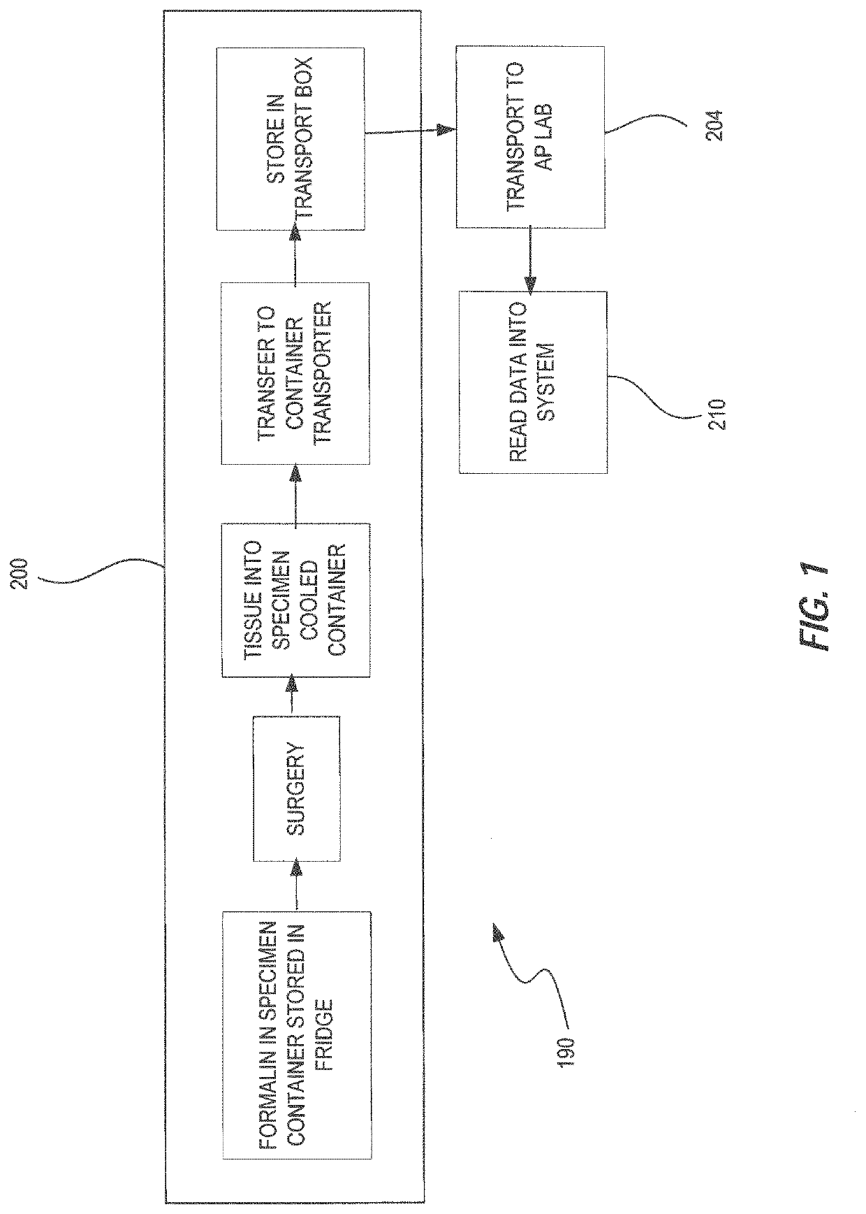
[0011]The disclosed integrated solution therefore can, in certain embodiments, result in 1) superior formalin fixation results for a broader range of biomarker-based diagnostics, including novel biomarker preservation previously only possible in fresh frozen specimens; 2) a processing time that is compatible with standard fixation protocols (depending on tissue type); 3) preservation of volatile biomarkers for up to 14 days prior to complete processing; 4) optionally, fully electronic tracking establishing a chain of custody that can be readily accessed from digital tablets and / or uploaded for cloud computing; 5) optionally, a real-time recording of multiple conditions that can be used to gauge whether additional measures will be necessary at destination to ensure optimal processing, including staining.
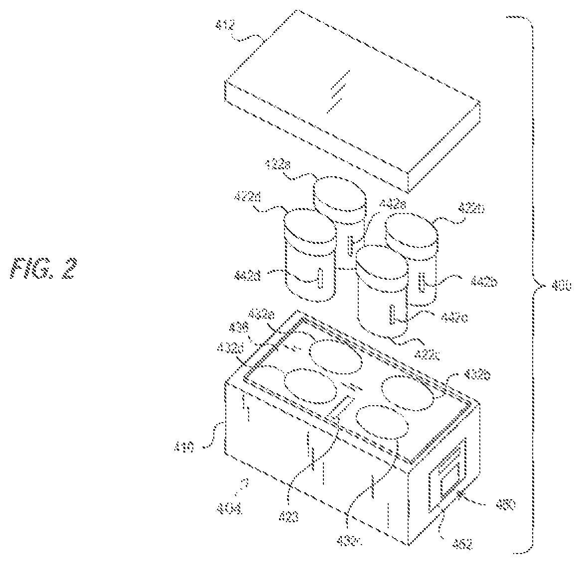
[0088]In one embodiment, the portable transporter system can include where cassette clip holds the standard histology cassette to ease reading of at least one code on the standard histology cassette.

Problems solved by technology

This also can lead to tissue that is either under exposed or over exposed by liquid fixatives.
For example, if formalin has not diffused properly through the tissue samples, outer regions of the tissue samples exposed to formalin may be over-fixed and interior regions of the tissue samples not exposed to formalin may be under-fixed, resulting in very poor tissue morphology.
In under-fixed tissue, subsequent exposure to ethanol often shrinks the cellular structures and condenses nuclei since the tissues will not have the chance to form a proper cross-linked lattice.
When under-fixed tissue is stained, such as with hematoxylin and eosin (H&E), many white spaces may be observed between the cells and tissue structures, nuclei may be condensed, and samples may appear pink and unbalanced with the hematoxylin stain.
Tissues that have been over-fixed typically do not work well for subsequent immunohistochemical (IHC) processes.
As a result, the optimal antigen retrieval conditions for these tissues do not work properly and therefore the tissue samples appear to be under stained.
Unfortunately, the tissue samples tend to exhibit over-fixed outer regions and under-fixed inner regions.
It may be difficult to identify such over-fixation and under-fixation upon arrival to the laboratory, thereby making it difficult to properly prepare the samples for examination.
The workers at the laboratory may not know when to remove the tissue samples from the formalin or other fixative.
If the specimen is left in the formalin an excess length of time, diagnosis may be delayed and the specimen may be over-fixed.
This under-fixation and over-fixation often results in poor preservation of biological molecules, tissue morphology, and / or post-translational modification signals.
It is not possible to ascertain what conditions the tissue sample in the container was exposed to or for how long.
There are several problems with current best-practice histology sample, collection, and transport methodologies.
Current room-temperature fixation protocols do not preserve biomarkers sufficiently.
Tissue storage after removal is not sufficiently standardized with respect to biomarker preservation; this can result in some specimens even being left without formalin over the weekend in a refrigerator.
The lack of rigid adherence to set protocols across institutions and sites often results in widely varying degrees of histomolecular degradation.
This compromises downstream processing, especially in samples subjected to a variety of biomarker-based diagnostics.
These results in the tissue often floating up to the surface, or adhered to the side of the wall of the container, thus compromising the amount of contact each sample has with the fixative fluid, the perfusion of which is critical for proper preservation and subsequent processing of the sample.
Additionally, variations in ambient temperature during transport are known to affect efficacy of fixative.
None of the above systems / devices solve the fixation and other problems mentioned above.
Real-time tracking of the computer specimen is paper-based and cannot provide an up-to-the-minute accounting of temperature, location and transport time.
The end result of these challenges is questionable sample quality (biomarker preservation may not correctly reflect the original state of the tissue) and lack of traceability during processing.
These are unacceptable risks when dealing with oncology patients.

Method used


Image

  • Transporter systems, assemblies and associated methods for transporting tissue samples
  • Transporter systems, assemblies and associated methods for transporting tissue samples
  • Transporter systems, assemblies and associated methods for transporting tissue samples

Examples


embodiment 1

2. The transporter system of embodiment 1 wherein the reagent is a tissue fixation reagent.

3. The transporter system of embodiment 1 further comprising a tissue sample cassette for retaining one or more tissue samples therein and wherein the specimen container is configured to retain and hold the tissue sample cassette in the reagent.

4. The transporter system of embodiment 1, wherein the monitoring system measures the temperature of the reagent and / or tissue sample in the specimen container over a period of time.

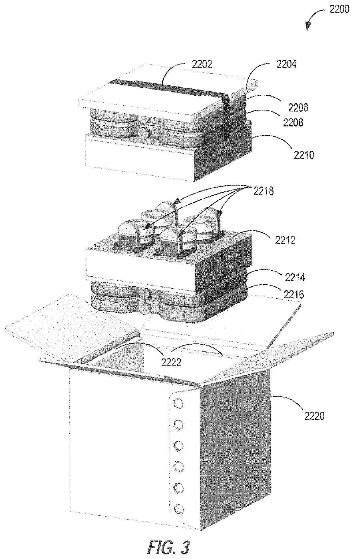
5. The transporter system of embodiment 1, further including an insulator and / or a cooling element capable of maintaining the volume of the formalin-based fixative solution at a temperature in the range of 0° C. to 15° C. for at least 2 hours under shipping conditions when the tissue sample transport and storage assembly is assembled.

6. The transporter system of embodiment 1, wherein the monitoring system is physically coupled to the carrier assembly.

7. The transporter syste...

embodiment 4

10. The transporter system of embodiment 4, wherein the specimen container comprises a temperature sensor wherein said sensor transmits the temperature of the reagent contained therein to the monitoring system.

11. The transport system of embodiment 1, wherein the specimen container further comprises a radio frequency identification (RFID) tag and the monitoring system further comprises a RFID reader.

12. The transport system of embodiment 1, further comprising a transport container for transporting the specimen container, carrier assembly and monitoring system wherein the transport container comprises an ice bath.

13. The transport system of embodiment 1, further comprising a transport container for transporting the specimen container, carrier assembly and monitoring system, wherein the transport container is configured to maintain the temperature of a specimen container therein to from about 0° C. to about 15° C. for up to 14 days.

14. The transporter system of embodiment 1, further c...

embodiment 16

17. The apparatus of embodiment 16, wherein the sensor assembly of each of the plurality of carrier assemblies comprises a temperature sensor positioned within the specimen container, wherein the temperature sensor includes a transmitter that outputs a temperature signal to a receiver located in the carrier assembly holding that specimen container.


PUM

PropertyMeasurementUnit
temperatureaaaaaaaaaa
freezing pointaaaaaaaaaa
temperatureaaaaaaaaaa

Abstract

A tissue sample that has been removed from a subject can be properly fixed for evaluation using the disclosed transporter assembly for carrying a tissue sample and method for fixing an unfixed tissue sample. In one embodiment, the disclosed assembly includes a transport container, a fixative in the transport container, and a cooling device that reduces and / or maintains the temperature of the fixative to perform a pre-soaking process at a temperature of less than about 7° C. The pre-soaking process can, for example, be performed during sample transport or during extended periods of storage, such as over a weekend.

Description

CROSS-REFERENCE TO RELATED APPLICATIONS[0001]This is a continuation of International Patent Application No. PCT / EP / 2016 / 051489, filed Jan. 26, 2016, the benefit of which is claimed. Benefit is further claimed to each of U.S. Provisional Patent Application No. 62 / 107,874, filed Jan. 26, 2015, and U.S. Provisional Patent Application No. 62 / 108,184, filed Jan. 27, 2015. The contents of all of these related applications are incorporated by reference herein.FIELD[0002]The present disclosure relates generally to transporter systems, assemblies and associated methods for transporting tissue samples. More specifically, the present disclosure relates to transporter systems and transporters for monitoring and / or analyzing the condition of tissue samples during transportation to improve the preservation of cellular activity.BACKGROUND[0003]Preservation of tissues removed by surgical procedures is a topic of great importance. After removal of a tissue sample from a subject, the tissue sample is...

Claims


Application Information

Patent Timeline
Patent Type & Authority Patents(United States)
IPC IPC(8): B01L3/00G01N35/10G01N1/31B01L9/06B01L7/04
CPCB01L3/502B01L9/06G01N1/31G01N35/10B01L3/50825B01L7/04B01L2200/026B01L2200/147B01L2200/18B01L2300/024B01L2300/047B01L2300/0609B01L2300/1855B01L2300/1877B01L2300/1883B01L2300/1894G01N2001/315
Inventor CRUM, NATHANKELLER, TIMOTHYLAING, ALASTAIROTTER, MICHAELPETRE, VINNIEPIGMAN, ELIZABETHSHINGLER, TAYLORSTANISLAW, STACEYSTEVENS, BENJAMIN
Owner VENTANA MEDICAL SYST INC
Learn More