
Mode of inducing renal transplant rejection on animals and its manufacturing approach

a technology of kidney transplant and manufacturing method, applied in the field of simplified model of kidney transplant in animals, can solve the problems of acute and chronic renal transplant rejection of patients, high risk of kidney transplantation, system bottleneck process, etc., and achieve the effect of inducing renal transplant rejection effectively

Active Publication Date: 2020-11-17
VETERANS GEN HOSPITAL TAIPEI +2
View PDF1 Cites 0 Cited by
  • Summary
  • Abstract
  • Description
  • Claims
  • Application Information

AI Technical Summary

Benefits of technology

This method effectively simulates transplant rejection, reducing animal mortality and improving reproducibility by directly inducing immune rejection in the renal cortex, facilitating the development of new immunosuppressive agents and understanding underlying mechanisms.

Problems solved by technology

Kidney transplantation is the best treatment for renal failure, but the risk of kidney transplantation is very high.
Therefore, the foreign kidney would lose its function and then result in necrosis.
As far as the surgery and therapy are concerned, kidney transplantation is not too complicated; however, how to control recipient's own immune system is the bottleneck process.
Nevertheless, the patients still have acute and chronic renal transplant rejection after administration of traditional immunosuppressive agents.
However, this method is likely to cause intraoperative or postoperative bleeding after vascular anastomosis.
The survival rate was low because the transplanted organ would be easily wrapped by mesentery and lose its function.
In addition, the renal transplantation in the previous literature is co-culturing the renal tubular epithelial cell lines with allogeneic lymphocytes in a cell model, but this model has high variability and cannot be applied to organisms.
Besides, the usage of skin allergy testing is different from the animal model of organ transplant rejection.
Therefore, it is necessary and urgent to improve the traditional kidney transplant procedure which is time-consuming with high animal mortality and lack of reproducibility and stability.

Method used


Image

  • Mode of inducing renal transplant rejection on animals and its manufacturing approach
  • Mode of inducing renal transplant rejection on animals and its manufacturing approach
  • Mode of inducing renal transplant rejection on animals and its manufacturing approach

Examples


example 1

and Culturing an Activated T Lymphocyte Platform In Vitro

[0028]Culturing Activated T-Lymphocytes In Vitro

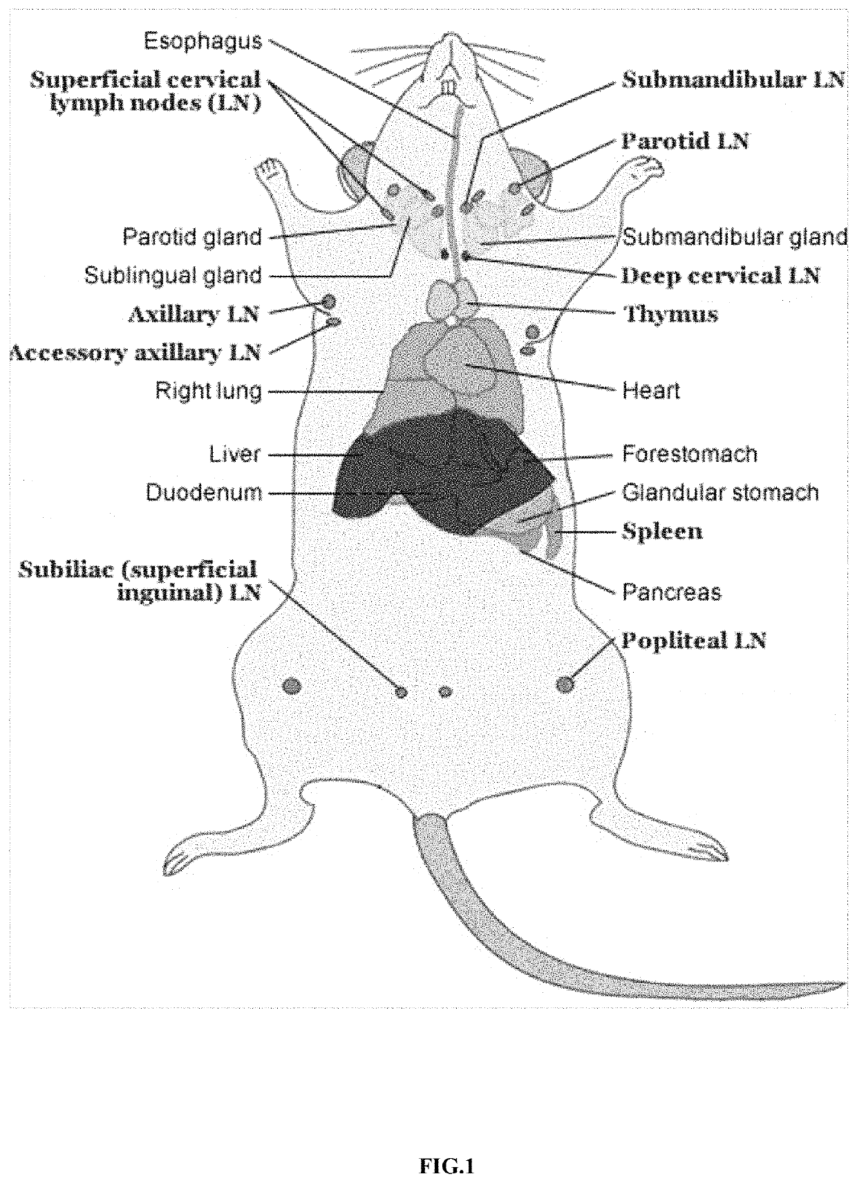
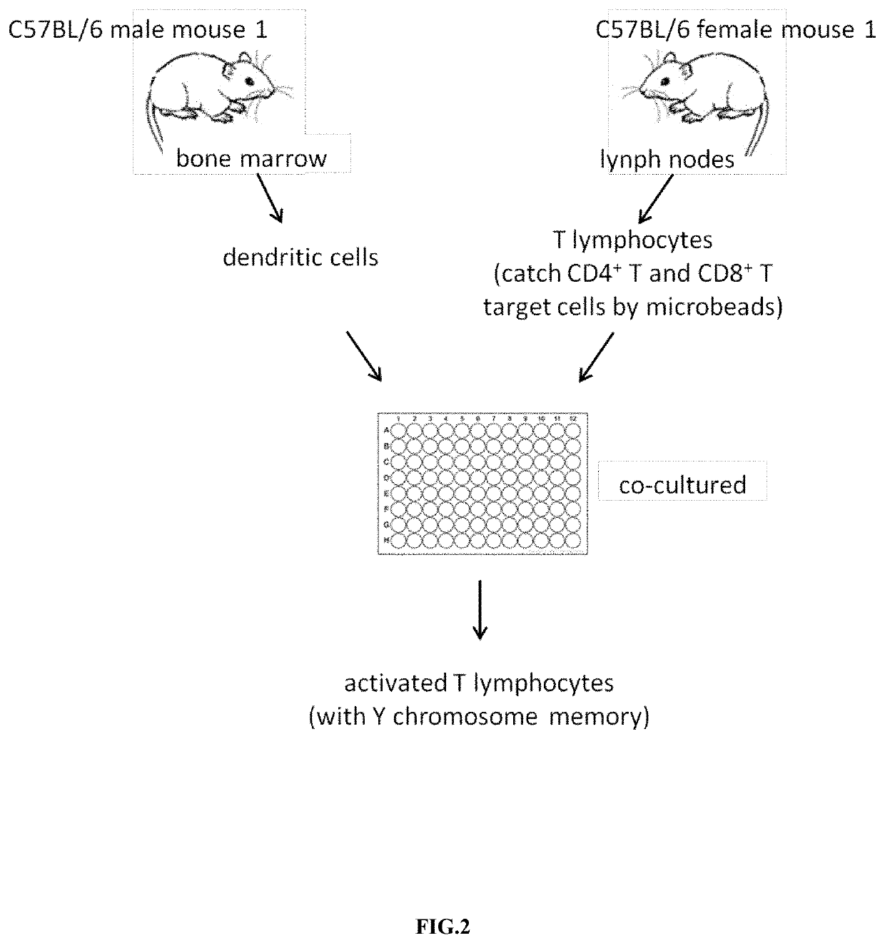
[0029]The dendritic cells of the male mouse C57BL / 6 were rushed out of the bone marrow (as shown in FIG. 1), and the dendritic cells were cultured in 6 ml Roswell Park Memorial Institute (RPMI) medium and granulocyte-macrophage colony-stimulating factor (GM-CSF) (20 ng / mL) for 9 days, and dendritic cells were stimulated with 100 ng / ml lipopolysaccharide (LPS) on the 9th day for about 24 hours; The lymph nodes of the mouse C57BL / 6 were found out, and the T lymphocytes were obtained (as shown in FIG. 1). CD4+ and CD8+ T cells were selected from these T lymphocytes by microbeads. Then, the dendritic cells and selected CD4+ and CD8+ T cells were co-cultured for 13 days to obtained the activated T lymphocytes (with Y chromosome memory) in vitro (FIG. 2).

example 2

tation of Cultured T Lymphocytes In Vitro

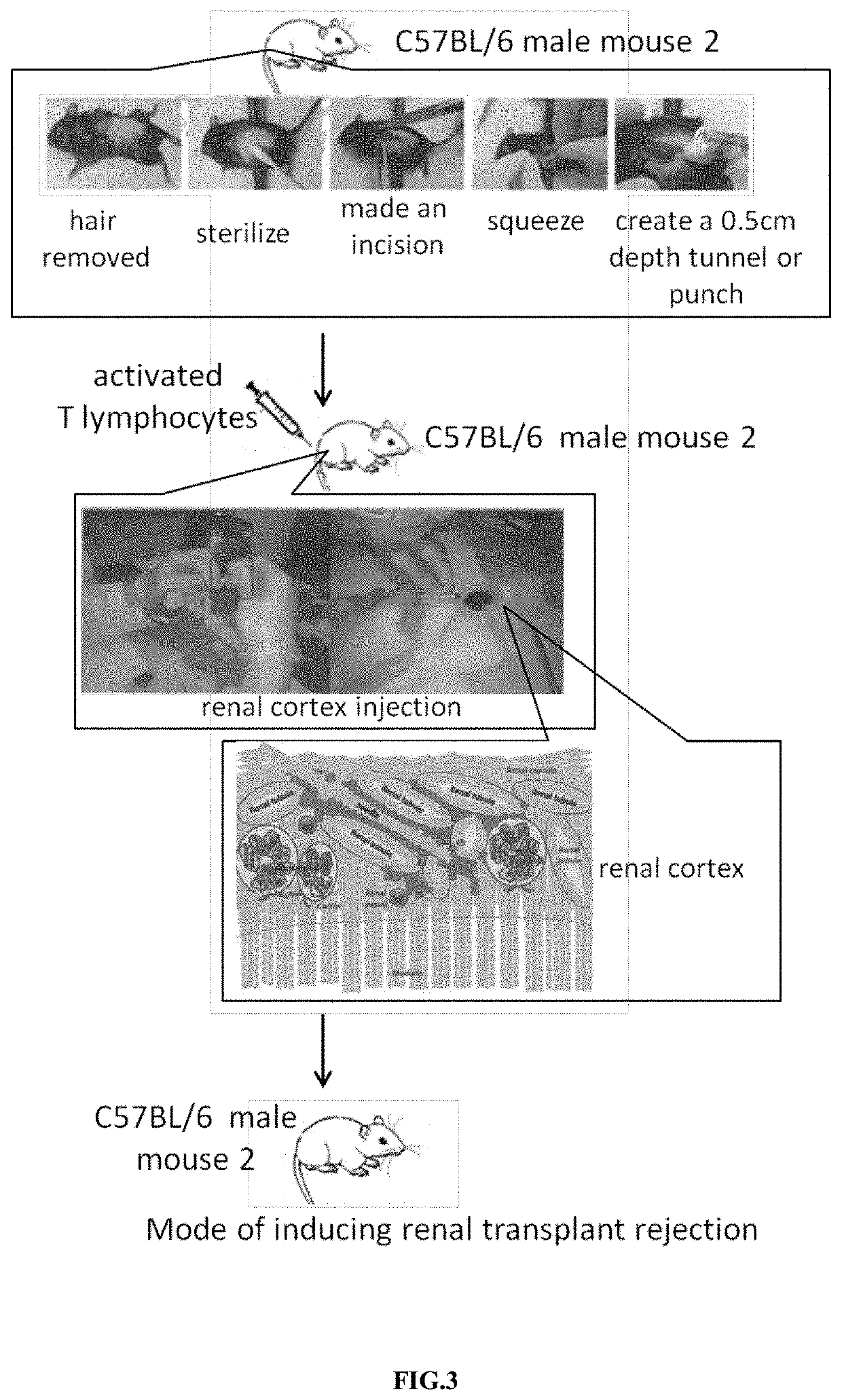
[0030]In order to facilitate the subsequent administration of activated T lymphocytes, we created a tunnel or pouch of the renal cortex in another C57BL / 6 male mouse. The surgical process for the manufacture of tunnels or pouches in renal cortex was shown in FIG. 3. The bilateral costal and hypochondriac region of the 6-10 weeks old mice were used for surgery. First, the hair was removed and sterilized. Then we created an opening near the kidney and squeezed out the kidneys with a sterile glove. Next, a needle No. 31 was used to make a 0.2 cm incision, and we injected the mouse kidney cortex along the long diameter to make a 0.5 cm depth tunnel or pouch to facilitate subsequent administration of activated T cells in vivo. 1×10⁶ to 3×10⁶ activated mouse T lymphocytes were counted in a cell counter, and these cells were injected into the tunnel or pouch made in above-mentioned renal cortex of another male mouse. The opening was closed by a supe...

example 3

ation Results from Tissue Slices

[0031]FIG. 5 showed the results of the hematoxylin and eosin (H & E) stain after the mouse kidney was sliced. The phosphate buffer solution (PBS) was injected into the tunnel or pouch of the renal cortex in the control group. 1×10⁶ activated T cells were injected into the tunnel or pouch of the renal cortex in the experimental group. From 0 to 14th day after injection, the kidney slices from the sacrificed mouse in the control group and in the experimental group were stained. The slices were observed under a 400-fold optical microscope with a scale of 50 μm. The arrows indicated that the renal tubules were not only structurally damaged by severe mononuclear infiltration, but also was found having renal tubular dilatation, renal tubular destruction, and extensive interstitial inflammatory cell infiltration. The results showed continuously pathological change in renal tissue both in control group and experimental group on day 0-day 14. There was no diff...


PUM

PropertyMeasurementUnit
depthaaaaaaaaaa
diameteraaaaaaaaaa
diameteraaaaaaaaaa

Abstract

The invention is a simplified model on kidney rejection in animals and its setting approach. The method involves isolating dendritic cells from male mice and isolating T cells from female mice and then using the naive T cells in vitro to culture concurrently with male dendritic cells. Those activated T cells from female origin were injected into renal cortex in male mice for the purpose of attacking renal cortex. The animal model simulates the renal transplant rejection method to enable effective induction of renal transplant immune reaction.

Description

CROSS-REFERENCE TO RELATED APPLICATIONS[0001]This application claims priority to Taiwan Application Serial No. 106115825, filed in May 12, 2017, the content of which is hereby incorporated by reference in its entirety.FIELD OF THE INVENTION[0002]The present invention is related to a simplified model on kidney rejection in animals and a method of creating the animal model, which comprises: isolating dendritic cells from male mice; isolating T cells from female mice; activating these dendritic cells in vitro; administrating these activated T cells (the activated T cells have a function of Y chromosome memory on the rodent cells in order to attack) into the renal cortex, to let the activated T cells attack the renal cortex of the male mice, in order to induce the renal transplant rejection effectively.BACKGROUND OF THE INVENTION[0003]Kidney transplantation is the best treatment for renal failure, but the risk of kidney transplantation is very high. When a solid organ was transplanted i...

Claims


Application Information

Patent Timeline
Patent Type & Authority Patents(United States)
IPC IPC(8): A61K49/00C12N5/0783A01K67/027
CPCA01K67/0271C12N5/0636C12N5/0638C12N5/0637A61K49/0008C12N2502/1114A01K2267/0325A01K2227/105C12N2502/1121A01K2207/12A61K39/4615A61K39/4611A61K39/4622A61K39/46434A61K2239/38A61K39/4621
Inventor TARNG, DER-CHENGCHEN, NIEN-JUNGWENG, SHUO-CHUN
Owner VETERANS GEN HOSPITAL TAIPEI
Learn More