Method for identification, selection and analysis of tumour cells

a tumour cell and selection technology, applied in the field of tumour cell identification, selection and analysis, can solve the problems of low sensitivity and specificity, widespread dosage of psa, poor sensitivity and specificity, and non-negligible costs for the health system, etc., to achieve high diagnostic and predictive reliability, high purity, and high accuracy.

Active Publication Date: 2019-03-19
MENARINI SILICON BIOSYSTEMS SPA
View PDF202 Cites 8 Cited by
  • Summary
  • Abstract
  • Description
  • Claims
  • Application Information

AI Technical Summary

Benefits of technology

[0022]Consequently, an aim of the present invention is to provide a method for identification, isolation, and subsequent analysis of circulating or disseminated rare cells obtained by means of a sampling of a non-invasive nature that will overcome the drawbacks of the known art described previously.
[0025]In particular, according to a preferential embodiment of the present invention the step of isolation of the rare cells is performed according to a technology developed by the present applicant and based upon the use of a silicon microchip that integrates several hundreds of thousands of electrodes of micrometric dimensions by manipulating individual cells so as to isolate the cells of interest with a unitary purity rendering them available for molecular analysis. In this way, the method according to the invention provides cells with a high purity, is highly automated for the most delicate part of the process (isolation of individual cells), and enables implementation of analytical techniques of high diagnostic and predictive reliability. In the light of what has been described previously, there will emerge clearly the innovative scope, at a diagnostic and therapeutic level, of the present invention, which enables improvement of the oncological treatment in the different stages of the course of the illness, rendering possible both a non-invasive early diagnosis having a level of reliability comparable with a biopsy, and a sensitive and accurate follow-up in the course of the treatment with specific cancer drugs and / or in the post-operative stage in order to follow the evolution of the illness and the quality of the response to the clinical treatments, both in the metastatic stage and in the adjuvant stage.

Problems solved by technology

Consequently, their sensitivity and specificity are not particularly high.
In the case, considered by way of example, of prostate cancer, one of the most widespread forms of cancer in the male population (approximately one quarter of all the cases of cancer, 300,000 new cases a year in Europe), screening on the basis of serological parameters (dosages of PSA) is widespread, but has a poor sensitivity and specificity.
More invasive procedures, such as digital rectal examination can at times complete the clinical picture, but more frequently it is necessary to carry out a prostatic biopsy to have a more accurate diagnosis, in an escalation of invasiveness that results in a growing discomfort for the patient and in non-negligible costs for the health system.
Since they are difficult to identify and eliminate, it is frequently impossible to treat all the metastases successfully.
Consequently, since there has been demonstrated, in the case of breast cancer, cancer of the colon, and cancer of the prostate, a correlation between the number of CTCs and the increased risk of unfavourable outcome of the illness, it appears evident that the possibility of isolating them and, by means of molecular analysis, obtaining a complete informative picture therefrom having clinical importance, would have very significant diagnostic repercussions.
13(5), 441-53 (2008)) that the over-expression of HER2, which can be detected in a single DTC but not in the primary tumour, proves predictive of a very high risk of death.
At the current state of the art, for isolation of circulating and disseminated tumour cells there are, however, available approaches of a chiefly analytical nature that are very complex and laborious, which, however, have limited yields and generate specimens distinguished by a low degree of purity.
Consequently, said specimens of CTCs or DTCs are not compatible with some of the most refined and reliable diagnostic procedures, such as sequencing.
The limited purity, in fact, determines a high risk of false positives and false negatives caused by wrong calls in the bases of the sequence.
However, said methods have led to levels of purity on average of 50%-60%, which do not prove sufficient for carrying out, downstream, analyses for which the purity of the specimen is crucial, such as, for example, the evaluation of copy-number variations, microsatellite instability, loss of heterozygosity (LoH), gene expression in which a better signal-to-noise (S / N) ratio is fundamental for diagnostic applications, or identification of new drugs, or again in the sector of research into cancer-initiating cells.
Similar considerations can be extended to other methods the enormous diagnostic potential of which has been highlighted, which, however, cannot be concretely implemented since the known procedures of enrichment of biological specimens and isolation of a population of rare cells therein do not enable sufficient levels of purity to be obtained, if not following upon a heavy, complex and costly analytical workload that cannot, consequently, find application in medical practice.
Also in this perspective, however, the methodologies of enrichment, identification and isolation of rare cells in the blood, or in an some biological fluid, do not enable a sufficient purity to be obtained to guarantee that the subsequent analysis will be reliable (exclusion of false positives, false negatives, etc.) and, in some cases, that they can even materially be conducted (for example, for the cases in which, since the purity is low, the signal-to-noise ratio is too low to obtain a valid reading).

Method used


Image

  • Method for identification, selection and analysis of tumour cells
  • Method for identification, selection and analysis of tumour cells
  • Method for identification, selection and analysis of tumour cells

Examples


example 1

[0175]Non-invasive evaluation of mutations (for example, in the K-RAS gene) in cancer patients.

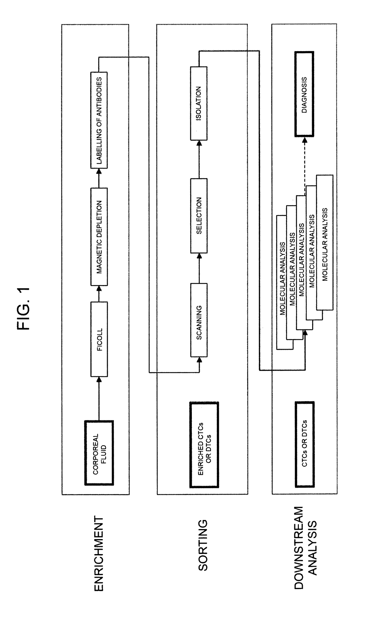
[0176]Described in what follows is the case of isolation of CTCs from peripheral bloodflow of patients affected by metastatic Colon-Rectal Cancer (mCRC).

[0177]A specimen of 7.5 ml of peripheral bloodflow was drawn from the patient in Vacutainer tubes with EDTA anticoagulant (Beckton Dickinson). The specimen was analysed with Coulter Counter (Beckman Coulter) and presented 42×106 leukocytes (WBCs) and 43.95×10⁹ erythrocytes (RBCs). The PBMCs were then isolated via centrifugation (in Ficoll 1077). The PBMCs recovered (16.5×10⁶ on the basis of the count with Coulter Counter) were washed in PBS with BSA and EDTA (Running Buffer, Miltenyi) and were selected via depletion with magnetic microbeads conjugated to anti-CD45 and anti-GPA antibodies (Miltenyi) according to the instructions of the manufacturer. The resulting cells (negative fraction for CD45 and GPA) were fixated with 2% PFA in PBS for...

example 2

[0191]Evaluation on CTCs of deletions (for example, in prostate cancer) via single-cell whole-genome amplification and CGH array. In this case, there is advantageously performed isolation of the single cells to be recovered in different wells. In this way, the analysis takes into account the heterogeneity of the population, and supplies not only the average, but multiple signals regarding each cell so that it is easier to identify mutations present only in some of the tumour cells.

example 3

[0192]Non-invasive identification of the original tissue in CUP tumours, via genetic profile and / or profile of expression of the CTCs.


PUM

PropertyMeasurementUnit
volumeaaaaaaaaaa
first wavelengthaaaaaaaaaa
third wavelengthaaaaaaaaaa

Abstract

The present invention relates to a method for the diagnosis of tumoural conditions and / or of the corresponding state of advance, wherein a specimen of an organic fluid taken from the patient and having a high probability of containing at least one circulating tumor cell (CTC) or a disseminated tumor cell (CTD) is enriched in a population of CTCs or CTDs. According to the invention, at least one type of CTCs or CTDs is isolated by selecting individually single cells in a microfluidic device so to constitute a diagnostic specimen having a purity of at least 90%. On the specimen thus obtained there is subsequently performed a molecular analysis such as to highlight a characteristic thereof suited to enabling diagnosis.

Description

CROSS-REFERENCE TO RELATED APPLICATIONS[0001]This is the U.S. national phase application of International Application No. PCT / IB2009 / 007316, filed Nov. 4, 2009, which claims the benefit of Italian Patent Application No. TO2008A 000814, filed Nov. 4, 2008.INCORPORATION BE REFERENCE OF MATERIAL SUBMITTED ELECTRONICALLY[0002]This application incorporates by reference in its entirety a computer-readable nucleotide / amino acid sequence listing identified as follows: One 607 byte ASCII (text) file name “46247_SeqListing.txt,” created Jan. 5, 2015.IN THE SEQUENCE LISTING[0003]Please introduce the computer-readable form of the Sequence Listing submitted herewith (filename: 46247_SeqListing.txt; Size: 607 bytes, Created: Jan. 5, 2015) as part of the application of the above-identified application as filed.FIELD OF THE ART[0004]The present invention relates, in general, to diagnostic methods based upon identification, isolation, and subsequent analysis of circulating or disseminated rare cells...

Claims


Application Information

Patent Timeline
Patent Type & Authority Patents(United States)
IPC IPC(8): G01N33/50G02B21/36B03C5/00G01N33/53
CPCG01N33/5023B03C5/005G01N33/5302G02B21/36G01N33/5091G01N2800/7028G01N33/574G01N33/52G01N1/28C12N5/0693G01N27/00G01N15/12C12Q1/6886C12N5/0081
Inventor MANARESI, NICOLMEDORO, GIANNIGIORGINI, GIUSEPPE
Owner MENARINI SILICON BIOSYSTEMS SPA
Why Eureka
Try Eureka