
Device for monitoring pressure in renal pelvis

A monitoring device and internal pressure technology, applied in medical science, urethroscopy, diagnosis, etc., can solve problems such as blood infection of patients

Pending Publication Date: 2022-07-29
THE THIRD MEDICAL CENT OF THE CHINESE PEOPLES LIBERATION ARMY GENERAL HOSPITAL
View PDF1 Cites 0 Cited by
  • Summary
  • Abstract
  • Description
  • Claims
  • Application Information

AI Technical Summary

Problems solved by technology

[0003] However, it is necessary to ensure that the pressure in the renal pelvis is kept within a reasonable range during the process of filling liquids such as water, normal saline or medicinal liquid. May cause serious bloodstream infection in patients

Method used


Image

  • Device for monitoring pressure in renal pelvis
  • Device for monitoring pressure in renal pelvis
  • Device for monitoring pressure in renal pelvis

Examples


Embodiment Construction

[0016] The present invention will be described in further detail below with reference to the accompanying drawings and embodiments. Wherein the same parts are denoted by the same reference numerals. It should be noted that the words "front", "rear", "left", "right", "upper" and "lower" used in the following description refer to directions in the drawings, and the words "bottom" and "top" "Face", "inner" and "outer" refer to directions toward or away from the geometric center of a particular part, respectively.

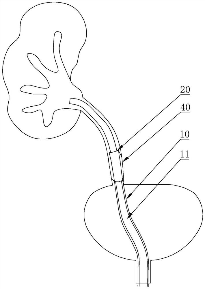
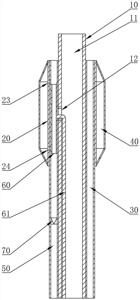
[0017] like figure 1 and figure 2 As shown, a pressure monitoring device in the renal pelvis includes a flexible ureteroscope 10, an operation channel 11 is opened in the flexible ureteroscope 10, and the flexible ureteroscope 10 is used to enter the renal pelvis from the ureter. A pressure detection collar 20 is arranged around the upper part, a pressure measurement channel 21 is opened on the pressure detection collar 20, a pressure measurement pipeline 30 is arr...


PUM


Abstract

The invention relates to a pelvis internal pressure monitoring device which comprises a flexible ureteroscope, an operation channel is formed in the flexible ureteroscope, the flexible ureteroscope is used for entering a pelvis from a ureter, a pressure detection lantern ring is arranged on the annular outer side wall of the flexible ureteroscope in a surrounding mode, a pressure measurement channel is formed in the pressure detection lantern ring, and the pressure measurement channel is communicated with the operation channel. A pressure measuring pipeline is arranged in the pressure measuring channel, one end of the pressure measuring pipeline is flush with the pressure measuring channel, the other end of the pressure measuring pipeline extends outwards and is connected with a pressure detector, and a fixing bag is arranged on the annular outer side wall of the pressure detecting lantern ring in a surrounding mode and used for fixing the position of the ureter. In conclusion, according to the technical scheme, the flexible ureteroscope has the advantages that the flexible ureteroscope is consistent with an existing flexible surgical scope and has the functions of illumination, lithotripsy, liquid injection and the like, and the pressure detection ring on the flexible ureteroscope can detect the pressure of the pelvis after the liquid is injected; and then the pressure in the pelvis is further adjusted by adjusting the water injection speed and the water injection amount through the flexible ureteroscope.

Description

technical field [0001] The present invention relates to the technical field of medical auxiliary equipment, and more particularly, to a pressure monitoring device in the renal pelvis. Background technique [0002] Flexible ureteroscopic lithotripsy is a treatment method for those patients in the kidney and ureter who cannot be crushed by traditional extracorporeal lithotripsy, or patients with kidney problems who cannot accept lithotripsy therapy and some huge kidney stones. . During lithotripsy surgery or ureteroscopy, liquids such as water, normal saline or medicinal solution need to be continuously poured into the renal pelvis and ureter to open the renal pelvis and ureter to open up the space, so that the flexible ureteroscope and optical fiber can be inserted into the ureter and the ureter. Laser lithotripsy in the renal pelvis. The invention patent with the publication number CN112674696A discloses a flexible ureteroscope, which includes a scope body and an operating...

Claims


Application Information

Patent Timeline
IPC IPC(8): A61B5/20A61B1/307
CPCA61B5/201A61B1/307A61B5/6867
Inventor 唐露
Owner THE THIRD MEDICAL CENT OF THE CHINESE PEOPLES LIBERATION ARMY GENERAL HOSPITAL
Learn More