Bionic drug-loaded nanoparticles for specific targeting pulsed electric field ablation postoperative inflammatory region and preparation method thereof
A pulsed electric field and drug-loaded nanotechnology, applied in the field of biomimetic drug-loaded nanoparticles and preparation, can solve the problems of high cost, complicated preparation process, and expensive activation steps, and achieve low cost, high safety, and medical transformation prospects. The effect of short preparation cycle
- Summary
- Abstract
- Description
- Claims
- Application Information
AI Technical Summary
Problems solved by technology
Method used
Image
Examples
Embodiment 1
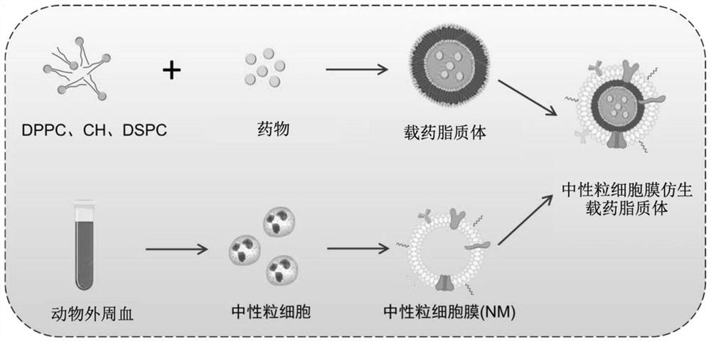
[0036] figure 1 It is a schematic flowchart of a method for preparing biomimetic drug-loaded nanoparticles specifically targeting the inflammatory area after pulsed electric field ablation provided by an embodiment. The preparation method of the biomimetic drug-loaded nanoparticles specifically targeting the inflammatory area after pulsed electric field ablation provided in this example includes the following steps:
[0037]Choose mice or rats. In this example, mice are used. After the mice are anesthetized with 5% chloral hydrate, blood is taken from the heart in an EP tube added with heparin. Take a 50ml centrifuge tube, carefully add mouse peripheral blood neutrophil separation solution according to the kit instructions, then carefully add blood samples, and centrifuge at 500g for 25min. After centrifugation, carefully absorb the neutrophil layer of the lower layer of cells, then add 3 times the volume of red blood cell lysate, mix gently by pipetting, lyse for 10 minutes,...
Embodiment 2
[0041] The difference between this example and Example 1 is that DPPC, CH, and DSPC are completely dissolved in chloroform at a molar ratio of 5:2:1, and ultrasonicated in a water bath at 60°C for a period of time.
Embodiment 3
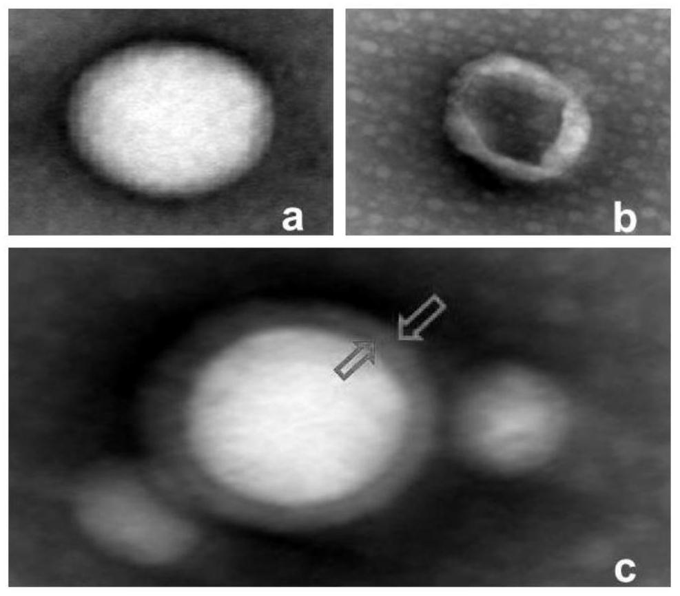
[0043] The drug-loaded liposomes, neutrophil membranes and neutrophil membrane biomimetic drug-loaded nanoparticles obtained in Example 1 were detected by transmission electron microscopy. The results of transmission electron microscopy are shown in figure 2 .
[0044] The results showed that the electron micrograph of the drug-loaded liposome was figure 2 In a, it can be seen that liposomes are round and uniform in particle size, and the dispersion system is good; the electron microscope image of neutrophil cell membrane is figure 2 In b, the double-layer cell membrane structure can be seen; the electron microscope image of the neutrophil membrane biomimetic drug-loaded nanoparticles is figure 2 In c, it can be seen that the round drug-loaded nanoparticles are wrapped in the double cell membrane.
PUM
| Property | Measurement | Unit |
|---|---|---|
| particle diameter | aaaaa | aaaaa |
Abstract
Description
Claims
Application Information
 - R&D
- Intellectual Property
- Life Sciences
- Materials
- Tech Scout
- Unparalleled Data Quality
- Higher Quality Content
- 60% Fewer Hallucinations
Browse by: Latest US Patents, China's latest patents, Technical Efficacy Thesaurus, Application Domain, Technology Topic, Popular Technical Reports.
© 2025 PatSnap. All rights reserved.Legal|Privacy policy|Modern Slavery Act Transparency Statement|Sitemap|About US| Contact US: help@patsnap.com


