
Surface-enhanced Raman scattering detection kit for detecting tumor micro nucleic acid markers as well as preparation method and application of surface-enhanced Raman scattering detection kit

A surface-enhanced Raman and detection kit technology, which is applied in the field of functional nanomaterials and biological detection, can solve the problems of non-specific false positive results, extensive restrictions, convenient application, and high requirements for primer design, so as to improve specificity and accuracy , highlight the application advantages, the effect of significant technical advantages

Pending Publication Date: 2022-04-29
NANJING UNIV OF POSTS & TELECOMM
View PDF0 Cites 1 Cited by
  • Summary
  • Abstract
  • Description
  • Claims
  • Application Information

AI Technical Summary

Problems solved by technology

[0003] Traditional miRNA detection methods include Northern blotting, microarray, and real-time polymerase chain reaction (RT-PCR), among which PCR-based detection technology is the main detection method. However, PCR technology has high requirements for primer design, and the amplification process It is easy to produce non-specificity and lead to false positive results, the detection is time-consuming, usually takes more than 2 hours, the operation is complicated, and it needs to be completed by professional technicians in a specialized laboratory
These problems limit its wide and convenient application

Method used


Image

  • Surface-enhanced Raman scattering detection kit for detecting tumor micro nucleic acid markers as well as preparation method and application of surface-enhanced Raman scattering detection kit
  • Surface-enhanced Raman scattering detection kit for detecting tumor micro nucleic acid markers as well as preparation method and application of surface-enhanced Raman scattering detection kit
  • Surface-enhanced Raman scattering detection kit for detecting tumor micro nucleic acid markers as well as preparation method and application of surface-enhanced Raman scattering detection kit

Examples


Embodiment 1

[0092] Example 1 Preparation of Surface Enhanced Raman Scattering (SERS) Detection Kit for Tumor Micronucleic Acid Marker Detection

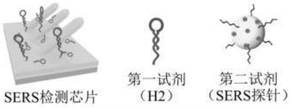
[0093] The kit consists of three parts, such as figure 1 Shown: SERS detection chip, first reagent and second reagent (ie SERS probe).

[0094] 1. Preparation of SERS detection chip and investigation of reaction conditions

[0095] 1. Preparation of SERS detection chip

[0096] (1) Prepare the silver nanorod array and rinse it with ultrapure water several times;

[0097] (2) After annealing the hairpin-type DNA single-strand H1 (heat at 95°C for 5-10 minutes, then cool down to 25°C in an ice-water bath), take the hairpin-type DNA single-strand H1 and TCEP solution (tricarboxyethylphosphine Solution) mixed at a molar ratio of 1:100 to 1:1000 and placed in a constant temperature mixer at 25°C for reaction;

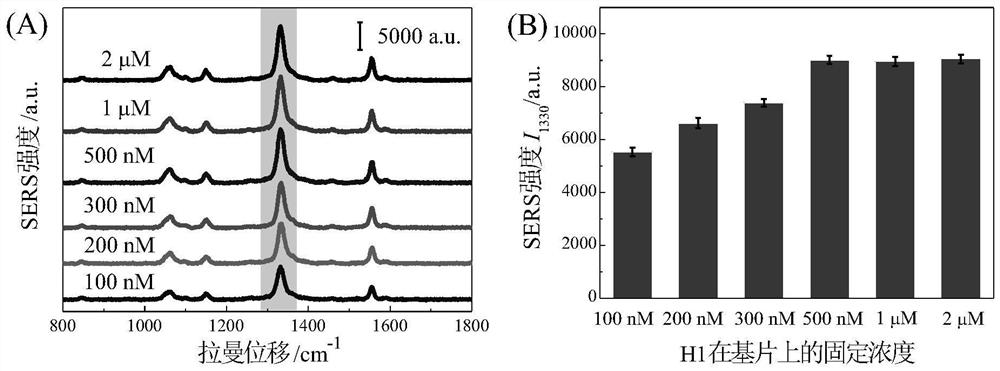
[0098] (3) Co-cultivate the silver nanorod array and 20 μL 100nM or 500nM hairpin DNA single-stranded H1 solution (cultivation conditions...

Embodiment 2

[0122] Example 2 Working curve and detection limit of miRNA-199a-3p detected by SERS detection kit

[0123] Mix 2 μL of 10 μM first reagent, 15 μL of second reagent, and 2 μL of sample solutions containing different concentrations of target miRNA-199a-3p (10aM-1nM), drop onto the surface of the SERS detection chip, and mix in a constant temperature mixer at 25°C and 300rpm After incubation in medium for 50 minutes, the wells were washed several times with reaction buffer (TM buffer) and ultrapure water in sequence. After natural air-drying, perform SERS test on the SERS detection chip (Raman test conditions: scanning time 1s, laser power 1%, objective lens magnification 20x, accumulation times 1 time, excitation light wavelength 785nm), and obtain the SERS spectrum and its characteristic signal intensity Value, take the logarithm of the target miRNA-199a-3p concentration as the abscissa, and use the characteristic peak intensity value of the SERS probe as the ordinate to make ...

Embodiment 3

[0124] Embodiment 3 SERS detection kit detects the working curve and detection limit of miRNA-100

[0125] Mix 2 μL of 10 μM first reagent, 15 μL of second reagent and 2 μL of sample solutions containing different concentrations of target miRNA-100 (10aM-1nM), drop onto the surface of the SERS detection chip, and incubate in a constant temperature mixer at 25°C and 300rpm After 50 minutes, the wells were washed several times with reaction buffer (TM buffer) and ultrapure water in sequence. After natural air-drying, perform SERS test on the SERS detection chip (Raman test conditions: scanning time 1s, laser power 1%, objective lens magnification 20x, accumulation times 1 time, excitation light wavelength 785nm), and obtain the SERS spectrum and its characteristic signal intensity Value, the logarithm of the target miRNA-100 concentration is taken as the abscissa, and the characteristic peak intensity value of the SERS probe is used as the ordinate to make a working curve, and t...


PUM


Abstract

The invention discloses a surface-enhanced Raman scattering detection kit for detecting small tumor nucleic acid markers for a non-disease diagnosis purpose as well as a preparation method and application of the surface-enhanced Raman scattering detection kit. The kit comprises an SERS detection chip, a first reagent and a second reagent. The SERS detection chip is a silver nanorod array substrate with the surface modified with a hairpin type DNA single chain H1, the first reagent is a hairpin type DNA single chain H2, the second reagent is an SERS probe, after the first reagent and the second reagent are added to the SERS detection chip at the same time, the single chain P of the probe on the second reagent is hybridized with the cohesive end of the H1-H2 double chain, and the SERS detection chip is a silver nanorod array substrate with the surface modified with the hairpin type DNA single chain H1. The SERS probe is captured on the SERS detection chip (the base sequence of the nucleic acid probe is adjustable), the detection of the small nucleic acid marker of the gastric cancer is realized by testing the Raman signal of the SERS probe on the SERS detection chip, the integrated detection of multiple nucleic acid biomarkers in complex environments such as serum and the like can be realized, and the detection specificity and accuracy are effectively improved.

Description

technical field [0001] The invention belongs to the field of functional nanomaterials and biological detection, and in particular relates to a surface-enhanced Raman scattering (SERS) detection kit for detection of small tumor nucleic acid markers and a preparation method and application thereof. Background technique [0002] In recent years, the incidence of tumors in my country has been increasing year by year and tends to be younger. It is the second leading disease after cardiovascular and cerebrovascular diseases. Tumors usually have an insidious onset, and early detection, early diagnosis, and early treatment are important measures to improve the survival rate. Therefore, breakthroughs in detection technology and methodology are urgently needed. Taking gastric cancer as an example, gastric cancer is one of the most common cancers in the world, which seriously threatens the life and health of the public. Gastric cancer is reported to be the fourth most common cancer a...

Claims


Application Information

Patent Timeline
IPC IPC(8): C12Q1/6886C12Q1/6837C12Q1/6806C12N15/11
CPCC12Q1/6886C12Q1/6837C12Q1/6806C12Q2600/178C12Q2525/301C12Q2563/157C12Q2563/137C12Q2565/632C12Q2545/114Y02A50/30
Inventor 宋春元汪联辉张晶晶
Owner NANJING UNIV OF POSTS & TELECOMM
Learn More