
3D bioprinted skin tissue model

A bioprinting and skin technology, applied in microorganisms, teaching models, tissue culture, etc., can solve problems such as not allowing controlled construction of in vitro models

Pending Publication Date: 2022-01-18
セルインクバイオプリンティングアーベー
View PDF10 Cites 1 Cited by
  • Summary
  • Abstract
  • Description
  • Claims
  • Application Information

AI Technical Summary

Problems solved by technology

However, these methods are labor intensive and do not allow controlled construction of in vitro models

Method used


Image

  • 3D bioprinted skin tissue model
  • 3D bioprinted skin tissue model
  • 3D bioprinted skin tissue model

Examples


Embodiment Construction

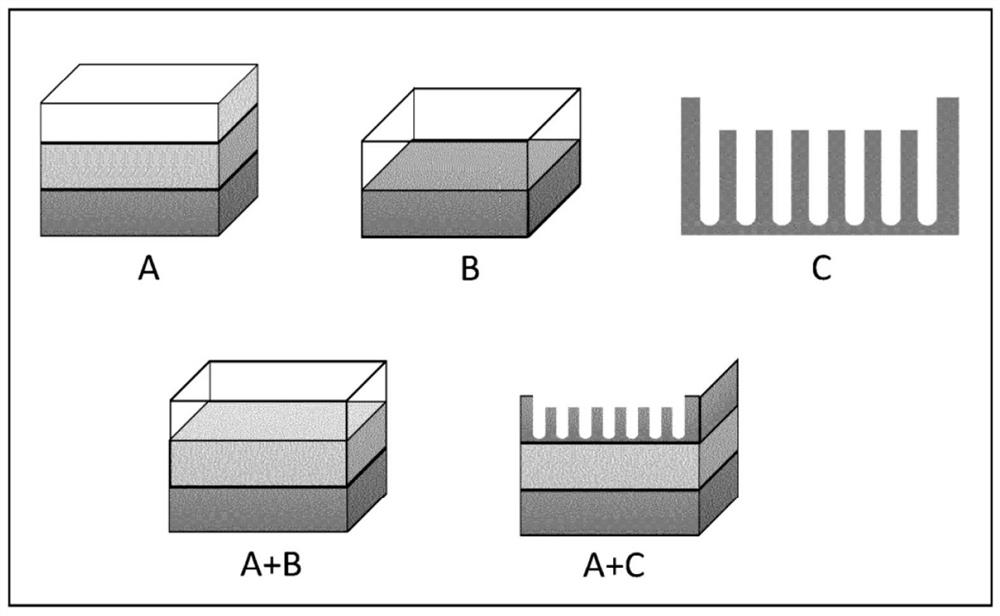
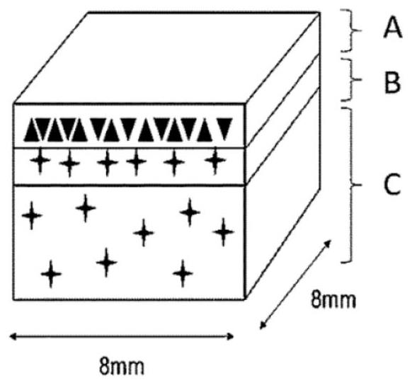
[0086] The present invention relates to a skin tissue model consisting of cells, biomolecules and bio-ink, for scientific research in the field of 3D modeling of skin tissue. Application of such a tissue model may be directed to cosmetic compound evaluation and / or discovery, medical device evaluation, skin care compound evaluation and / or discovery, drug evaluation and / or discovery, medical regeneration, Vigor tissue and / or cell recovery, photosensitivity testing, drugs and / or molecular compound absorption test, tissue engineering, toxicology studies, stimulating research, testing and allergen skin physiology and / or pathology. Cells, biomolecules and bio-ink deposition by a particular layered to simulate the natural distribution of natural skin cells and extracellular matrix, for both the inner and outer liner skin, such as skin, esophagus and urethra. When the skin tissue model constructed stable in culture, in a gas - liquid interface culture of the model to mimic the...


PUM


Abstract

The present invention relates to a 3D bioprinted skin tissue model, a method for providing said model and the use of said model. The 3D bioprinted skin tissue model comprises at least one bioink A, at least one cell type A, at least one factor A, wherein the bioink A comprises at least one biopolymer, a thickener, at least one extra-cellular matrix or a decellularized matrix, and optionally a photo initiator and / or cellular additions, the at least one cell type A is an epidermal, dermal and / or hypodermal cell or cell line, and the at least one factor A is a growth factor, protein and / or molecule thatstimulates altered or abnormal metabolism of cell type A.

Description

Technical field [0001] The present invention relates to the field of 3D printing the biological tissue, and in particular relates to a skin tissue model. Background technique [0002] In the field of in vitro skin model, the gold standard cell culture is cultured in a two-dimensional (2D) culture cells are allowed to form a confluent layer of the epidermis analog compartment. This may be carried out in 2D culture is covered with a layer of dermal fibroblasts feeder into the tissue culture plastic, or dermal fibroblast cells having a three-dimensional (3D) structure. Such 3D structures are generally molded, or is seeded with dermal fibroblasts scaffold. Since the human body tissues and cells are distributed in 3D space, and therefore these types of cell culture enhances the similarity of the natural tissue environment. However, these methods are labor-intensive, and does not allow a controlled in vitro Models. Therefore, development can provide highly relevant and standardized cel...

Claims


Application Information

Patent Timeline
Patent Type & Authority Applications(China)
IPC IPC(8): A61L27/38A61L27/44A61L27/60C12N5/00C12N5/071
CPCA61L27/44A61L27/60C12N5/0062C12N5/0697C12N2503/02C12N2503/04C12N2506/45C12N2513/00C12N2533/52C12N2533/54C12N2533/74B33Y70/00A61L27/3804G09B23/306B33Y80/00A61F2/105A61L27/20C12M21/08C12N5/0698B29C64/10B33Y10/00B33Y30/00B33Y70/10C12N2503/06
Inventor 伊莎贝拉·邦德松阿德尔·尔特达·纳姆罗雷德万赫克托·马丁尼兹埃里克·盖滕霍尔姆
Owner セルインクバイオプリンティングアーベー
Learn More