
Light-activated probe with organelle sequential imaging and double-color imaging functions and application thereof

A two-color imaging and photoactivation technology, applied in the field of photoactivated probes, can solve the problems of differences in intake, poor residence capacity and photostability, adverse mitochondrial morphological changes, etc., and achieve the effect of a simple method.

Pending Publication Date: 2022-01-14
SOUTH CHINA UNIV OF TECH
View PDF7 Cites 1 Cited by
  • Summary
  • Abstract
  • Description
  • Claims
  • Application Information

AI Technical Summary

Problems solved by technology

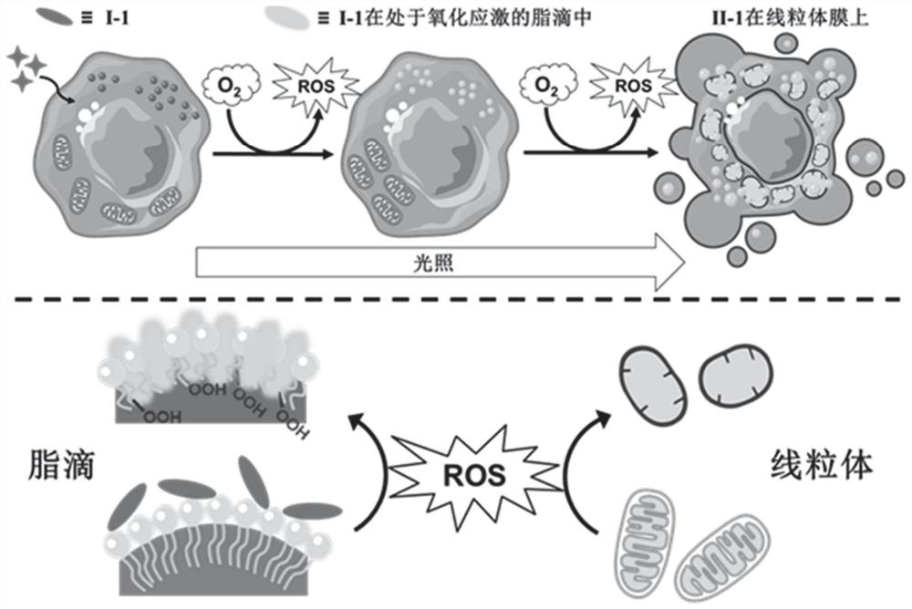
Therefore, in order to achieve simultaneous imaging of mitochondria and lipid droplets, researchers usually need to add at least two fluorescent dyes, but this method is severely limited by: (1) differences in the uptake of different dyes by cells; (2) tedious staining process; (3) overlapping of dye emission spectra, etc.
In addition, the existing lipid droplet and mitochondrial dyes are deficient: on the one hand, almost no lipid droplet dyes can directly identify lipid droplets under oxidative stress in a "fluorescent light-up" manner; on the other hand, commercial mitochondrial The dye has poor retention ability and photostability, which is not conducive to long-term monitoring of mitochondrial morphological changes

Method used


Image

  • Light-activated probe with organelle sequential imaging and double-color imaging functions and application thereof
  • Light-activated probe with organelle sequential imaging and double-color imaging functions and application thereof
  • Light-activated probe with organelle sequential imaging and double-color imaging functions and application thereof

Examples


Embodiment 1

[0054] Preparation of Compound I-1:

[0055] Weigh compound II-1 (28.3mg, 0.04mmol) in a reaction vessel, add 5mL methanol to it, and ultrasonically dissolve the solid completely; weigh sodium borohydride (15.5mg, 0.41mmol) and add it to the reaction solution, and Stir the reaction at room temperature under nitrogen atmosphere for half an hour; after the reaction is completed, use a rotary evaporator to depressurize the reaction solution to obtain the crude product to be treated; add 10mL ultrapure water to the crude product, ultrasonicate, and filter to obtain a brown solid product. The precipitate was dried in vacuo to obtain compound I-1 in ~8.5% yield.

[0056] The structure of compound I-1 is

[0057] The structure of compound II-1 is

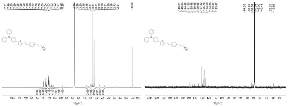
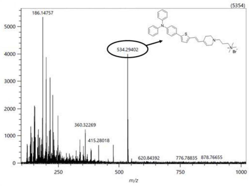
[0058] The chemical structure characterization data of the product are as follows:

[0059] 1 H NMR (MeOD, 400MHz): δ7.49(d, J=8.8Hz, 2H), 7.29(t, J 1 =15.6Hz,J 2 =8.4Hz, 4H), 7.22(d, J=8.0Hz, 2H), 7.19(d, J=3.6Hz, 2H), 7.07(d, J...

Embodiment 2

[0063] The characterization of compound I-1 and compound II-1 photophysical properties, as shown in Table 1:

[0064] Table 1 Characterization of photophysical properties of compound I-1 and compound II-1 in different solvents

[0065]

[0066] Figure 4 It is a characterization diagram of the photophysical properties of compound I-1; (A) the ultraviolet absorption spectrum of compound I-1, and its fluorescence emission spectrum (40μM) in phosphate buffer or 1,4-dioxane; (B) Fluorescence emission spectrum (40 μM) of compound I-1 in the mixed solution of 1,4-dioxane and water continuously increasing 1,4-dioxane content, and the excitation wavelength is 371nm; (C) Compound I-1 (10μM) in the mixed solution of 1,4-dioxane and water, the ratio change diagram of the fluorescence emission intensity at 463nm and the maximum emission wavelength change diagram with increasing 1,4-dioxane content ; (D) schematic diagram of LUMO and HOMO energy levels of compound I-1;

[0067] Fig...

Embodiment 3

[0073] Characterization of the photoactivation process of compound I-1:

[0074] Figure 7 It is a characterization diagram of the photoactivation process of compound I-1; (A) compound I-1 in liposome solution with light (40mW / cm 2 ) time-varying ultraviolet absorption spectrum (40μM); (B) compound I-1 in liposome solution with light (40mW / cm 2 ) time-varying fluorescence emission spectrum (40μM), the excitation wavelength is 371nm; (C) compound I-1 in liposome solution with light (40mW / cm 2 ) time-varying fluorescence emission spectrogram (40μM), the excitation wavelength is 488nm; (D) the absorption intensity of compound I-1 in liposome solution at 371nm and 481nm varies with light (40mW / cm 2 ) time ratio change figure; (E) Fluorescence emission intensity of compound I-1 in liposome solution at 445nm with light (40mW / cm 2 ) Ratio change figure of time, the excitation wavelength is 371nm; 2 ) time ratio diagram, the excitation wavelength is 488nm.

[0075] Such as Figu...


PUM


Abstract

The invention belongs to the technical field of light-activated fluorescent probes, and discloses a light-activated probe with organelle sequential imaging and double-color imaging functions and application thereof. The structure of the light-activated fluorescent probe is as shown in formula I, and R1-R4 are independent hydrogen; and R5 represents an alkylene carboxyl group, an alkylene ester group, or an alkylene ammonium salt group. The light-activated fluorescent probe is applied to fluorescence imaging of lipid droplets and mitochondria, in particular to sequential imaging and double-color imaging of lipid droplets and mitochondria in cells. The light-activated fluorescent probe is used for monitoring the interaction between lipid droplets and mitochondria under oxidative stress and monitoring the morphological change of the lipid droplets and / or mitochondria under oxidative stress. The light-activated fluorescent probe can selectively lighten lipid droplets in an oxidative stress state, in-situ light-activated products of the light-activated fluorescent probe in cells can specifically dye mitochondria, and the probe has the advantages of being good in light stability, high in mitochondria residence capacity, high in signal-to-noise ratio, large in Stokes shift and the like.

Description

technical field [0001] The invention belongs to the technical field of medical materials and relates to a light-activated probe, in particular to a light-activated probe with functions of organelle sequential imaging and two-color imaging and its application. The light-activated probe of the present invention is a tetrahydropyridine derivative substituted by triphenylamine thiophene vinyl, which is used for sequential imaging of different organelles and monitoring the interaction of different organelles under oxidative stress. Background technique [0002] Oxidative stress refers to a state of redox imbalance, which is closely related to the occurrence and development of many diseases, such as cancer, inflammation and neurodegenerative diseases. Usually, excessive reactive oxygen species (ROS) in cells can disrupt the intracellular redox balance, thereby inducing oxidative stress in cells. Studies have shown that organelles play an important role in ROS-induced oxidative st...

Claims


Application Information

Patent Timeline
IPC IPC(8): C07D409/06C09K11/06G01N21/64
CPCC07D409/06C09K11/06G01N21/6456C09K2211/1014C09K2211/1029C09K2211/1092Y02B20/00
Inventor 王迎军张子聪高蒙
Owner SOUTH CHINA UNIV OF TECH
Learn More