Application of MST4 related substances in preparation of medicine for treating neuroinflammation reaction after cerebral hemorrhage
A related substance, MST4 technology, applied in the direction of drug combination, neurological diseases, pharmaceutical formulations, etc., to achieve the effect of alleviating neurological deficits
- Summary
- Abstract
- Description
- Claims
- Application Information
AI Technical Summary
Problems solved by technology
Method used
Image
Examples
Embodiment 1
[0049] Embodiment 1, experimental design
[0050] 1. Experimental design: such as figure 1 As shown, mice were assigned to four independent experiments and all design details are listed.
[0051] Experiment 1: 30 mice were divided into sham operation group and 6h, 12h, 24h and 72h groups after cerebral hemorrhage. The levels of MST4 and NLRP3 were detected by western blot. Brain tissue was taken for immunofluorescence (IF) detection.
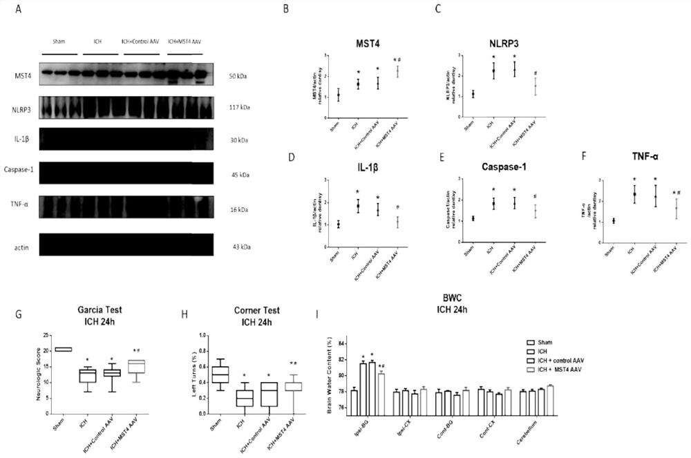
[0052] Experiment 2: Randomly divided into sham operation group, ICH group, ICH+control AAV group, ICH+MST4 AAV group, 60 each. Western blot detection of MST4, NLRP3, IL-1β, caspase-1 and TNF-α protein levels (n=6), brain tissue IF (n=3), neurological function test 24h after cerebral hemorrhage (n=15) and brain water content (n=6).
[0053] Experiment 3: 30 rats in each of the ICH group and the ICH+Hesperidin group. All mice were sacrificed 24 hours after cerebral hemorrhage, and western blot (n=6), BWC (n=6), neurological examination (n=1...
Embodiment 2
[0067] Example 2. Expression of MST4 and NLRP3 and their cellular localization after ICH
[0068] MST4 protein levels were significantly increased at 12 and 24 hours after ICH and peaked at 12 hours (*p figure 2 A, B). Elevated NLRP3 protein levels were observed at 12 and 24 hours after ICH, peaking at 24 hours (*p figure 2 A, C). Both MST4 and NLRP3 levels decreased with the peak. 24 hours after ICH was chosen as the observation time point for experiments 2, 3 and 4. Immunofluorescence of MST4 or NLRP3 with Iba1 was performed. Both MST4 and NLRP3 were expressed in microglia and increased after ICH ( figure 2 D).
Embodiment 3
[0069] Example 3, MST4 AAV inhibits the activation of NLRP3 inflammasome
[0070] The expressions of MST4, NLRP3 inflammasome components, IL-1β, and TNF-α increased after ICH (*p image 3 A-F). MST4 AAV attenuated caspase-1 activation, IL-1β and TNF-α secretion (#p image 3 A-F). MST4 AAV administration significantly improved neurological deficits (*p image 3 G-H). Consistent with neurological assessment, BWC was elevated in the ipsilateral hemisphere after ICH whereas MST4 AAV died BWC (*p image 3 I).
PUM
 Abstract
Description
Claims
Application Information
 - R&D
- Intellectual Property
- Life Sciences
- Materials
- Tech Scout
- Unparalleled Data Quality
- Higher Quality Content
- 60% Fewer Hallucinations
Browse by: Latest US Patents, China's latest patents, Technical Efficacy Thesaurus, Application Domain, Technology Topic, Popular Technical Reports.
© 2025 PatSnap. All rights reserved.Legal|Privacy policy|Modern Slavery Act Transparency Statement|Sitemap|About US| Contact US: help@patsnap.com


