Pathological image automatic classification method based on dyeing intensity matrix

An automatic classification and pathological image technology, applied in the field of medical image processing, can solve the problem of limited processing speed, and achieve the effects of high diagnostic accuracy, practicability and easy implementation.

Active Publication Date: 2021-10-22
ZHEJIANG LAB
View PDF3 Cites 4 Cited by
  • Summary
  • Abstract
  • Description
  • Claims
  • Application Information

AI Technical Summary

Problems solved by technology

[0005] In order to solve the technical problem that the accuracy and processing speed of the existing computer-aided diagnosis method based on pathological images are limited by the performance of the color normalization algorithm, the present invention proposes an automatic classification method for pathological images based on the staining intensity matrix

Method used


Image

  • Pathological image automatic classification method based on dyeing intensity matrix
  • Pathological image automatic classification method based on dyeing intensity matrix
  • Pathological image automatic classification method based on dyeing intensity matrix

Examples


Embodiment Construction

[0037] The present invention will be further described in detail below with reference to the accompanying drawings and specific embodiments, and the contents not described in detail belong to the prior art known to those skilled in the art.

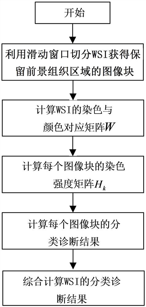
[0038]This embodiment takes the diagnosis and classification of lung adenocarcinoma and lung squamous cell carcinoma as an example. The image block classification network uses ResNet50, and the random forest algorithm is used to synthesize the classification and diagnosis results of each image block. The training data has been marked by professional radiologists. 200 full-section digital pathological images of cancer types and regions, including 100 lung adenocarcinoma and 100 lung squamous cell carcinoma. The diagnosis and classification of lung adenocarcinoma and lung squamous cell carcinoma using the proposed method for automatic classification of pathological images based on staining intensity matrix includes the following steps (such ...


PUM


Abstract

The invention discloses a pathological image automatic classification method based on a dyeing intensity matrix. The method comprises directly extracting a dyeing intensity matrix irrelevant to a dyeing agent ratio, a dyeing platform, a scanning platform and some human factors in a pathological image as classified feature information. A normalized dyed image does not need to be recovered, so that all impurity-free information related to diagnosis is reserved, and meanwhile, the phenomenon that the diagnosis effect of an existing pathological image computer-aided diagnosis method based on a traditional color normalization method is changed along with the change of a selected standard pathological section is avoided, and errors caused by the fact that the dyed image needs to be recovered are avoided. According to the invention, the diagnosis precision is higher, the diagnosis effect is more stable, meanwhile, diagnosis of pathological images can be achieved in a shorter time, implementation is easy, and practicability is higher.

Description

technical field [0001] The invention relates to the technical field of medical image processing, in particular to an automatic classification method for pathological images based on a staining intensity matrix. Background technique [0002] Histopathological examination is a pathomorphological method for analyzing and diagnosing diseases by examining pathological changes in tissues. It is currently the most accurate method for diagnosing cancer. Whole Slide Image (WSI) is a high-magnification large-scale digital image that can be displayed, transmitted and processed by a computer formed by scanning histopathological sections with a dedicated microscopic imaging system. Although digital pathological slice imaging technology has been promoted and applied in major medical institutions, the current diagnosis and analysis of WSI still requires doctors to make repeated observations under different scales of magnifying glass to obtain the diagnosis results. The diagnosis results de...

Claims


Application Information

Patent Timeline
Patent Type & Authority Applications(China)
IPC IPC(8): G06T7/00G06K9/46G06K9/62G16H70/60G06N3/04
CPCG06T7/0012G16H70/60G06N3/045G06F18/241G06F18/214G06T2207/30024G06T2207/20021G06T2207/20081G06T2207/20084G06T2207/10024G06T2207/10056G06T7/194G06T7/11G06T7/90G06T7/0002
Inventor 朱闻韬薛梦凡李少杰杨德富
Owner ZHEJIANG LAB
Why Eureka
Try Eureka