
Reagent composition and application thereof for one-step screening and/or diagnosis of clonal diseases

A composition and reagent technology, applied in the field of blood disease detection, can solve the problems of low detection efficiency of rare tumors and precancerous lesions, heavy workload of the two-step method, and large individual differences in schemes, so as to facilitate the accumulation of data and experience, The effect of speeding up reporting time and reducing workload

Active Publication Date: 2021-11-02
信纳克(北京)生化标志物检测医学研究有限责任公司
View PDF8 Cites 0 Cited by
  • Summary
  • Abstract
  • Description
  • Claims
  • Application Information

AI Technical Summary

Problems solved by technology

[0005] In order to solve this problem, researchers have made research explorations, including the schemes launched by the European Flow Consortium (euroflow) since 2012, but the euroflow scheme still has the following defects: 1. It is a screening based on the first step The two-step method of investigation, which leads to the very important judgment of the first step, if the personnel make a wrong judgment in the first step, it will lead to the wrong choice of antibody in the second step; 2. The first step of the two-step method is relatively simple, not It may cover all disease screening, so it is easy to miss diagnosis; 3. The two-step method has a large workload and individual differences in the scheme, which is not conducive to the automation of pre-processing, automatic sample loading and other processes, as well as the development of multi-dimensional software analysis and artificial intelligence such as flowjo; 4. The focus is on the diagnosis of common hematological tumors such as acute leukemia and lymphoma. It does not involve metastatic cancer, and the detection efficiency of rare tumors and precancerous lesions is not high; 5. Most clinical patients are diagnosed with anemia, leukopenia, three It is difficult to solve the situation where there is no clinical information, and even if other tests are done, they cannot provide effective information.
[0006] After investigation, there is no more convenient and feasible method in the prior art that can be applied to flow cytometry screening for almost all clonal diseases (including tumors, AA, pure red AA, rare normal polymorphisms, and common precancerous lesions, immunodeficiency, etc.)

Method used

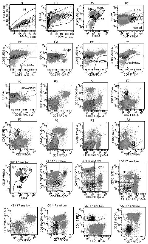
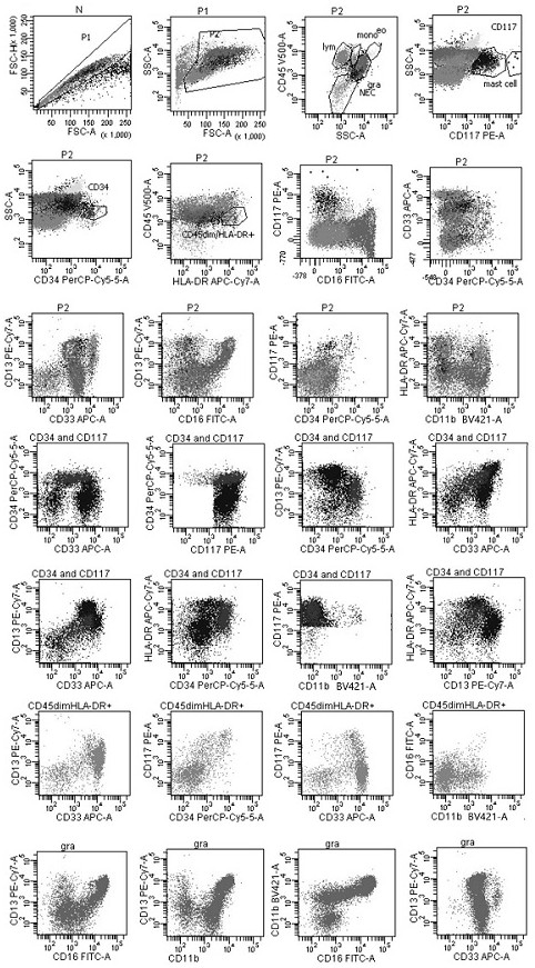
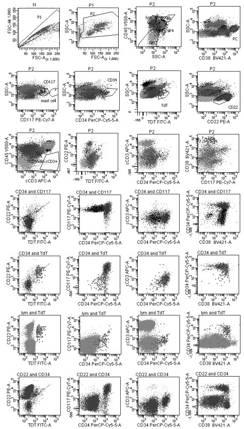

Image

  • Reagent composition and application thereof for one-step screening and/or diagnosis of clonal diseases
  • Reagent composition and application thereof for one-step screening and/or diagnosis of clonal diseases
  • Reagent composition and application thereof for one-step screening and/or diagnosis of clonal diseases

Examples


Embodiment 1

[0094] The preparation of embodiment 1 reagent

[0095] The antibody combination used in this example is,

[0096] The first group is: the first group of antibodies includes: anti-CD7 antibody, anti-CD117 antibody, anti-CD3 antibody, anti-CD4 antibody, anti-CD5 antibody, anti-CD8 antibody, anti-CD56 antibody, anti-CD45 antibody and anti-CD2 antibody, each antibody The fluorescein labeling sequence is FITC, PE, PerCP-Cy5.5, PE-Cy7, APC, APC-Cy7, BV421, V500 and BV605; take the above nine monoclonal antibody reagents in a volume ratio of 5:5:5:3 : 2:3:3:3:3 volume ratio mixed in the first container;

[0097] The second group is: anti-CD16 antibody, anti-CD117 antibody, anti-CD34 antibody, anti-CD13 antibody, anti-CD33 antibody, anti-HLA-DR antibody, anti-CD11b antibody and anti-CD45 antibody. The fluorescein labeling sequence of each antibody is FITC, PE, PerCP-Cy5.5, PE-Cy7, APC, APC-Cy7, BV421 and V500; the above eight monoclonal antibody reagents are mixed according to the ...

Embodiment 2

[0106] Example 2 Processing of Specimen

[0107] According to the results of cell counting, add heparin or EDTA anticoagulated bone marrow or peripheral blood samples into flow tube A, and ensure that the amount of cells added is about 2×10 6 According to Table 1, add 32 μl of nine kinds of cell membrane monoclonal antibody reagents labeled with different fluorescein to the flow tube, mix well with the cell suspension, incubate at room temperature and avoid light for 15 minutes, add 3ml 1× hemolysin, avoid Incubate with light for 10 minutes to lyse red blood cells, centrifuge at 1500rpm for 5 minutes to remove the supernatant, add 3ml of PBS to mix, centrifuge at 1500rpm for 5 minutes to remove the supernatant, add 0.5ml of PBS buffer to resuspend the cells, that is, the processed specimen, which can be used for On-board testing.

[0108] According to the results of cell counting, add heparin or EDTA anticoagulated bone marrow or peripheral blood samples into flow tube B, and...

Embodiment 3

[0114] Example 3 Detection of Specimen

[0115] The specimens processed according to the method of Example 2 were detected on the 3-laser 10-color FACS Canto plus flow cytometer of Becton Dickinson Company in the United States. After obtaining 1 million cells per tube (recommended at least 300,000), use diva 2.8 Software or other software such as Kaluza to analyze the data.

[0116] Among them, the flow cytometry gate is set in the following ways: ①Fixed gate: remove the adhesion cell gate, live cell gate, and blood cell gate in sequence; ②multi-marker combination gate: start from a single living cell, in order To prevent missing tumor cells, the gating and definition of all cells need to be carried out under parallel conditions with the blood cell gate; ③In the multi-marker combination gate, the common expression patterns and development patterns of various marker combinations are displayed, based on the normal Different cells, to find tumor cells; or naive T cells or naive ...


PUM

PropertyMeasurementUnit
Sensitivityaaaaaaaaaa

Abstract

The invention provides a reagent composition for one-step screening and / or diagnosis of clonal diseases and its application. The reagent composition includes eight groups of antibodies, which are compositions for flow cytometry detection that can be used for one-step screening and / or diagnosis of clonal diseases. When applied, a 5-tube parallel scheme is used, wherein the first group of antibodies, The second set of antibodies were used for different tube samples, the third set of antibodies and the sixth set of antibodies were used for the same tube samples, the fourth set of antibodies and the seventh set of antibodies were used for the same tube samples, the fifth set of antibodies and the eighth set of antibodies for samples in the same tube. The reagent composition of the present invention can be applied to one-step flow cytometry screening / diagnosis of various clonal diseases and other abnormalities, including: acute leukemia, mature lymphocytic tumor, chronic myeloid tumor, nocturnal paroxysmal nocturnal hemoglobin Urine, metastatic cancer, precancerous lesions, rare normal polymorphisms, aplastic anemia, abnormalities in other immune cell subsets, etc.

Description

technical field [0001] The invention relates to a reagent composition for one-step screening and / or diagnosis of clonal diseases and its application, belonging to the technical field of blood disease detection. Background technique [0002] The clonal disease here generally refers to a type of cell abnormality-related diseases, not limited to malignant tumors, but also includes a certain type of consistent cell proliferation caused by factors such as chronic infection and genetic abnormalities, such as nocturnal paroxysmal nocturnal hemoglobinuria (Paroxysmal Nocturnal Hemoglobinuria, PNH), aplastic anemia (AA), pure red AA, rare normal polymorphisms, common precancerous lesions, immune deficiency, etc. Malignant tumors have always been one of the important diseases that affect human health. The total incidence of malignant tumor clonal diseases such as acute leukemia (AL), lymphoma, metastatic cancer, chronic myeloid tumors, and PNH has reached 1 in 1,000 More than one, an...

Claims


Application Information

Patent Timeline
Patent Type & Authority Patents(China)
IPC IPC(8): G01N33/577G01N33/58G01N33/574G01N33/68G01N15/14
CPCG01N33/577G01N33/582G01N33/57492G01N33/57496G01N33/6893G01N15/14G01N2800/24G01N2800/34G01N2800/60G01N2800/22G01N15/1459G01N2015/1006G01N2015/1488G01N2015/1402G01N15/1429G01N2333/70596G01N33/56966G01N33/57426C07K16/2803C07K16/2887C07K16/289C07K16/2896
Inventor 王卉陈曼王爱先宫美维吴雪英甄军毅杜晴郭亚
Owner 信纳克(北京)生化标志物检测医学研究有限责任公司
Learn More