Drainage device used for medical oncology

A technology for internal medicine and tumors, which is applied in the direction of using tools for cleaning methods, suction devices, cleaning methods and appliances, etc. It can solve the problems of drainage tubes shaking or cheapness, and drainage tubes cannot be guaranteed, and achieve good sterilization effect.

Pending Publication Date: 2021-09-17
ANHUI PROVINCIAL CANCER HOSPITAL
View PDF0 Cites 0 Cited by
  • Summary
  • Abstract
  • Description
  • Claims
  • Application Information

AI Technical Summary

Problems solved by technology

Use the tip of a knife to cut a small incision in the skin where the needle enters, dilate the tube and dilate it, inject a pigtail catheter along the guide wire, withdraw the guide wire when all side holes are behind the pleural effusion, and leave the drainage tube In the thoracic cavity, the drainage tube is fixed with skin sutures, and a sterile drainage bag is connected externally, and the tube drainage is completed; but this method of fixing the drainage tube will make the drainage tube shake or be cheap when the patient moves. , cannot ensure that the drainage tube inserted into the patient's body does not deviate; therefore, we need a drainage device for medical oncology

Method used


Image

  • Drainage device used for medical oncology
  • Drainage device used for medical oncology
  • Drainage device used for medical oncology

Examples


Embodiment 1

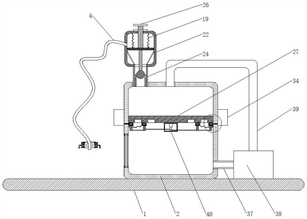
[0032] Embodiment 1: A third spring fixed to the inner wall of the processing box 2 is fixed on the sides of the two groups of fourth fixing rods 32 located at the edge part away from each other; the cleaning cotton can be restored to its original position by the restoring force of the third spring.

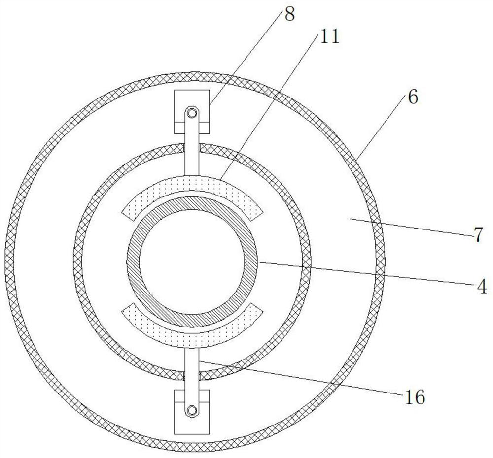
[0033] Working principle: when fixing the catheter 4, first place the fixing collar 6 at the incision where the patient is about to insert the catheter, then rotate the second trachea 14, and the second trachea 14 drives the suction cup 15 connected to it to rotate together, so that each One group of suction cups 15 can be in close contact with the patient's skin, and then the catheter 4 is inserted into the patient's body from the incision, and then the two groups of first fixed rods 16 are pulled, so that the two groups of first fixed rods 16 are close to each other, and the movement of the first fixed rods 16 drives them The connected clamping plates 11 move together, and the t...


PUM


Abstract

The invention belongs to the field of tumor medical instruments, and particularly relates to a drainage device used for the medical oncology. The drainage device comprises a bottom plate, wherein a treatment box is fixed at the top of the bottom plate; an anti-backflow mechanism is fixedly connected to the top of the treatment box; a catheter is fixedly connected to one side of the anti-backflow mechanism; a fixing mechanism is arranged on the outer ring of the catheter; the fixing mechanism comprises a fixing lantern ring of a hollow structure; a first fixing plate which has an annular structure and is coaxially arranged with the fixing lantern ring is arranged on the inner ring of the fixing lantern ring; two groups of first springs symmetrically distributed along the central axis of the first fixing plate are fixed between the first fixing plate and the inner wall of the bottom of the fixing lantern ring; two groups of pushing plates which have a trapezoidal structure and are symmetrically distributed along the central axis of the first fixing plate are fixed to the top of the first fixing plate; the top of each pushing plate is slidably connected with a first fixing rod which is in slidable sleeving with the fixing lantern ring and is of an L-shaped structure; and a clamping plate of an arc-shaped structure is fixed to the other end of each first fixing rod. By use of the drainage device disclosed by the invention, the catheter can be clamped and fixed, and the catheter is prevented from sliding out of the body.

Description

technical field [0001] The invention relates to the technical field of tumor medical devices, in particular to a drainage device used in oncology medicine. Background technique [0002] In the surgical resection of human tumors, there is often effusion in the tumor. In the traditional process of tumor effusion drainage, pleural effusion drainage can be divided into one-step method and two-step method under ultrasound. The two-step method is more commonly used in clinical practice. . The two-step method is to use an 18G-21G PTC needle to puncture the effusion according to the puncture point positioned before the ultrasound operation, and then pull out the needle core to see the liquid flow out, or use a syringe to aspirate and see the liquid, which proves that The puncture needle has entered the chest cavity. At this time, a guide wire can be inserted through the PTC needle. When the guide wire enters the pleural effusion through the needle tip to a sufficient depth, the gu...

Claims


Application Information

Patent Timeline
IPC IPC(8): A61M1/00A61M25/02A61L2/18A61L2/10B08B1/00B08B13/00
CPCA61M25/02A61L2/18A61L2/10B08B13/00B08B1/143
Inventor 秦汉林胡长路冯克海
Owner ANHUI PROVINCIAL CANCER HOSPITAL
Why Eureka
Try Eureka