Construction method of endothelial cell and pericyte co-culture model for researching tubulation
A technology of endothelial cells and construction methods, applied in the field of construction of endothelial cells and pericyte co-culture models, can solve the problems of unclear correlation of pericyte cancer, affecting the effectiveness of screening drugs targeting blood vessels, and unknown molecular mechanism, etc., to achieve Reduce the effect of animal models and clinical experimental tests, good social and economic value, and improve tumor vascular function
- Summary
- Abstract
- Description
- Claims
- Application Information
AI Technical Summary
Problems solved by technology
Method used
Image
Examples
Embodiment 1
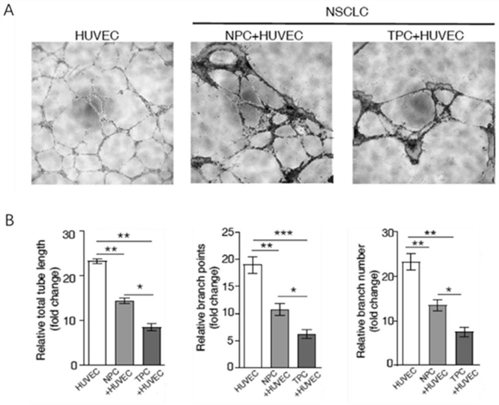
[0054] This example provides a method for co-culturing human endothelial cells and pericytes in vitro to study tube formation. All isolation and culture operations are carried out in a biological safety cabinet, which specifically includes the following steps:
[0055] (1) Rinse the human umbilical cord vein (from a hospital in Guangzhou) with normal saline until it is colorless, pour 37°C preheated trypsin into the human umbilical cord vein with a syringe, seal it, and incubate at 37°C for 15 minutes. Endothelial cells were insufflated into complete DMEM medium to terminate digestion.
[0056] (2) Centrifuge the cells at room temperature at 300g for 5 minutes, resuspend and disperse them into single cells with endothelial cell medium (ECM), and then place the cells in a 6-well plate at 37°C and 5% CO 2 Incubate for 24 hours. The suspended cells were sucked away to replace the medium, and the adherent growth cells were human umbilical vein endothelial cells (HUVEC).
[0057]...
Embodiment 2
[0068] The reagents in other steps during the tube formation process were the same as those in Example 1, except that the ratios of human umbilical vein endothelial cells (HUVEC) and TPC in step (10) were 5:1 and 20:1. After co-cultivating HUVEC and TPC cells, the reticular vascular structure can also be observed under a fluorescence microscope (the experimental results are similar to those in Example 1, so no additional figures are shown).
Embodiment 3
[0070] The reagents in the remaining steps during the tube formation process were the same as in Example 1, except that the final concentration of the CFSE dye in step (9) was 2 μM or 10 μM. After co-cultivating HUVEC and TPC cells, the reticular vascular structure can also be observed under a fluorescence microscope (the experimental results are similar to those in Example 1, so no additional figures are shown).
PUM
 Abstract
Description
Claims
Application Information
 - R&D
- Intellectual Property
- Life Sciences
- Materials
- Tech Scout
- Unparalleled Data Quality
- Higher Quality Content
- 60% Fewer Hallucinations
Browse by: Latest US Patents, China's latest patents, Technical Efficacy Thesaurus, Application Domain, Technology Topic, Popular Technical Reports.
© 2025 PatSnap. All rights reserved.Legal|Privacy policy|Modern Slavery Act Transparency Statement|Sitemap|About US| Contact US: help@patsnap.com


