Eureka AIR delivers breakthrough ideas for toughest innovation challenges, trusted by R&D personnel around the world.

Automatic white matter lesion quantitative analysis system and interpretation method

A technology for quantitative analysis of white matter in the brain, applied in the field of medical imaging, can solve problems such as time-consuming and labor-intensive, difficult to achieve accurate evaluation of disease curative effect evaluation, prognosis prediction, lack of quantitative analysis and information integration, etc.

Active Publication Date: 2021-07-06
WEST CHINA HOSPITAL SICHUAN UNIV
View PDF13 Cites 2 Cited by
  • Summary
  • Abstract
  • Description
  • Claims
  • Application Information

AI Technical Summary

Problems solved by technology

However, the imaging manifestations of these diseases are complex and have certain similarities. At present, the imaging of diseases mainly relies on the subjective experience and judgment of radiologists, lacks quantitative analysis and information integration, and lacks intuitive display of image descriptions.
At the same time, the different seniority and writing habits of doctors make the report content and terminology also different, resulting in the lack of standardized and standard report writing methods, which is not only time-consuming and labor-intensive, but also difficult to accurately judge the disease, which may delay patient treatment Maximize the role of imaging examinations in this type of disease
More importantly, many diseases in white matter lesions are chronic diseases, which require long-term follow-up and observation to evaluate the changes of the disease. The existing imaging reporting method only relies on human eye observation, which cannot objectively and accurately measure the volume of the lesion. It is difficult to achieve accurate evaluation in terms of follow-up, curative effect evaluation, and prognosis prediction.

Method used


Image

  • Automatic white matter lesion quantitative analysis system and interpretation method
  • Automatic white matter lesion quantitative analysis system and interpretation method
  • Automatic white matter lesion quantitative analysis system and interpretation method

Examples


Embodiment Construction

[0127] The present invention will be further elaborated below in conjunction with embodiment.

[0128] 1. Establish clinical information knowledge base module:

[0129] The scope of use of this structured report is the MRI examination of white matter lesions, the first step, to determine the age of the patient, for example, age: □ Under 65 years old (check) □65 to 75 years old □75 years and above; the second part determines whether the patient has vascular risk factors: □None (selected) □Hypertension □Hyperlipidemia □Diabetes □Smoking history □Obesity □Others (such as hypercoagulable state, vasculitis , migraine, etc.[])□unknown; the third step is to determine whether there is any other relevant clinical history: Others: [].

[0130] 2. Anatomical model map module

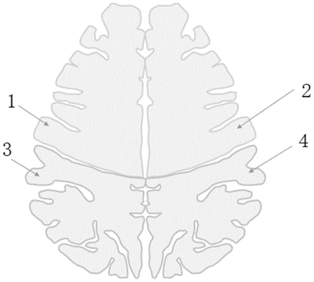
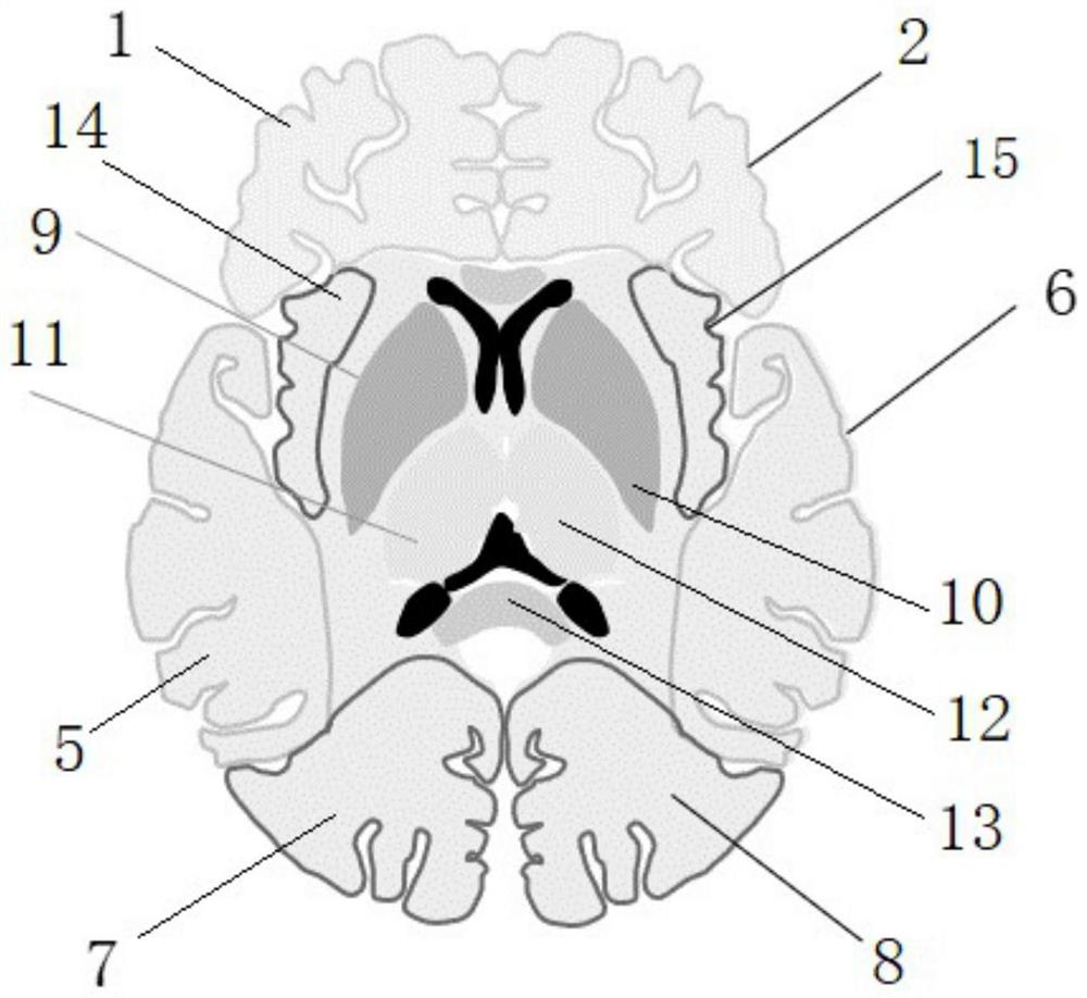
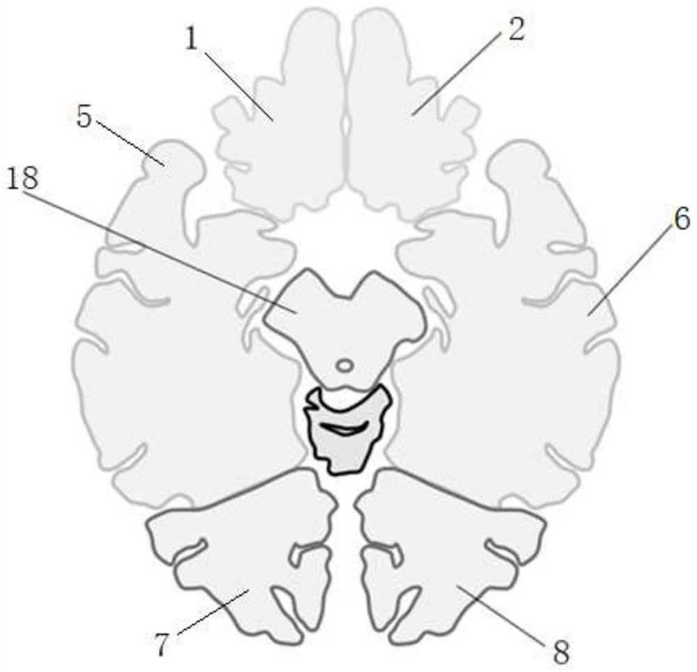
[0131] The computer displays the cross-sectional schematic diagram of each brain anatomical structure in the model map module. The radiologist uses the mouse to click on the distribution and location of the whit...


PUM


Abstract

The invention discloses an automatic quantitative analysis system and an interpretation method for white matter lesions, which utilize an artificial intelligence method to automatically and accurately sketch lesions, accurately calculate the volume of the white matter lesions and provide possibility for accurate evaluation of disease progression. Meanwhile, the invention provides the automatic interpretation system, multi-dimensional information such as anatomical positioning, morphological structure, lesion size, lesion occurrence time and bleeding of lesions is determined through human-computer interaction, lesion positioning visualization, image index quantification, report term standardization and operation interface friendliness are achieved, logic analysis is automatically made, disease judgment is accurately obtained, and inconformity between a conclusion and description and missing report of important conclusions are avoided.

Description

technical field [0001] The present application relates to the field of medical imaging, in particular to a method for automatic quantitative analysis of lesion volume and intelligent diagnosis of diseases based on multimodal MRI images of patients. Background technique [0002] The brain is the most complex organ of human beings and an important part of the nervous system. The brain is divided into three parts: gray matter, white matter, and cerebrospinal fluid. Vascular diseases, inflammatory diseases, demyelinating diseases and other diseases may cause lesions in the white matter of the brain, leading to limb fatigue, cognitive impairment, aphasia, epilepsy and other symptoms in patients. It is difficult to rely on clinical symptoms alone Accurate diagnosis of patients at an early stage of the disease. MRI (Magnetic resonance imaging, MRI) has high resolution of soft tissue and no radiation, and plays a vital role in the examination of neurological diseases, especially b...

Claims


Application Information

Patent Timeline
IPC IPC(8): G16H50/20G06T7/00G06T7/11G06N3/04G06N3/08
CPCG16H50/20G06T7/0012G06T7/11G06N3/08G06T2207/10088G06T2207/20081G06T2207/20084G06T2207/30016G06N3/045
Inventor 姚骊吕粟曾嘉欣胡娜李思燚张文静
Owner WEST CHINA HOSPITAL SICHUAN UNIV
Why Eureka
Eureka Blog
Learn More