
CT image segmentation method based on artificial neural network

An artificial neural network, CT image technology, applied in the field of medical CT image processing, can solve the problems of uneven grayscale, incomplete segmentation, and many noise points, and achieve the effect of facilitating subsequent processing, speeding up segmentation, and ensuring integrity.

Active Publication Date: 2021-06-25
SHANGHAI FIRST PEOPLES HOSPITAL
View PDF11 Cites 2 Cited by
  • Summary
  • Abstract
  • Description
  • Claims
  • Application Information

AI Technical Summary

Problems solved by technology

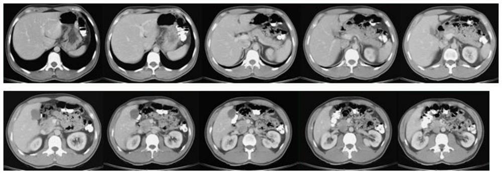
[0002] CT (Computed Tomography), that is, electronic computerized tomography, it uses precisely collimated X-ray beams and highly sensitive detectors to scan a certain part of the human body one by one, and the generated CT images can assist doctors Judgment and treatment require the professionalism and proficiency of doctors. Nowadays, convolutional neural networks have made remarkable achievements in the field of image segmentation, and have been applied to organ recognition and region detection in medical images. However, due to the partial Volume effect, gray level inhomogeneity, artifacts, and the proximity of gray levels between different soft tissues, for accurate extraction of the region of interest from the CT image, and further zoom-in analysis, there are incomplete segmentation and many noises

Method used


Image

  • CT image segmentation method based on artificial neural network
  • CT image segmentation method based on artificial neural network
  • CT image segmentation method based on artificial neural network

Examples


Embodiment Construction

[0028] The following will clearly and completely describe the technical solutions in the embodiments of the present invention with reference to the accompanying drawings in the embodiments of the present invention. Obviously, the described embodiments are only some, not all, embodiments of the present invention. Based on the embodiments of the present invention, all other embodiments obtained by persons of ordinary skill in the art without making creative efforts belong to the protection scope of the present invention.

[0029] It should be noted that when a component is said to be "fixed" to another component, it can be directly on the other component or there can also be an intervening component. When a component is said to be "connected" to another component, it may be directly connected to the other component or there may be intervening components at the same time. When a component is said to be "set on" another component, it may be set directly on the other component or t...


PUM


Abstract

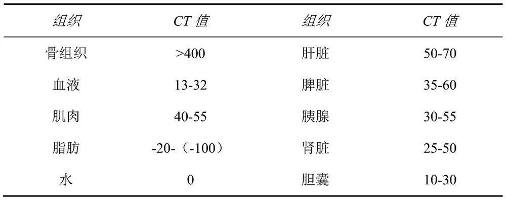
The invention relates to the technical field of medical CT image processing, and discloses a CT image segmentation method based on an artificial neural network, and the method comprises the following steps: carrying out the preprocessing of a CT image; segmenting the skeleton part, obtaining the outer contour of the abdominal cavity, and determining the number of faults; carrying out internal organ feature recognition in the outline of the skeleton, and extracting the outline of a single internal organ; processing, segmenting and storing the outline of the viscera in a classified mode; judging whether all viscera organs in the fault are segmented or not, if not, returning to reprocess, and otherwise, performing the next step; extracting a plurality of pieces of fault data of a certain viscera organ in a centralized manner according to requirements; According to the method, the skeleton part is firstly segmented, the area where the internal organs are located is rapidly determined, data processing of follow-up internal organ recognition and segmentation is reduced, invalid recognition is reduced, and the segmentation speed is increased; by setting the standard library, the segmented image data is detected, and the integrity of the segmented image is ensured.

Description

technical field [0001] The invention relates to the technical field of medical CT image processing, in particular to a CT image segmentation method based on an artificial neural network. Background technique [0002] CT (Computed Tomography), that is, electronic computerized tomography, it uses a precisely collimated X-ray beam and a highly sensitive detector to scan a certain part of the human body one by one, and the generated CT images can assist doctors Judgment and treatment require the professionalism and proficiency of doctors. Nowadays, convolutional neural networks have made remarkable achievements in the field of image segmentation, and have been applied to organ recognition and region detection in medical images. However, due to the partial Volumetric effects, gray level inhomogeneity, artifacts, and the proximity of gray levels between different soft tissues, for accurate extraction of the region of interest from the CT image, and further zoom-in analysis, there ...

Claims


Application Information

Patent Timeline
Patent Type & Authority Applications(China)
IPC IPC(8): G06T7/12G06T7/13G06T7/136G06K9/46G06T5/00
CPCG06T7/12G06T7/13G06T7/136G06T2207/10081G06T2207/20032G06T2207/20192G06T2207/30008G06V10/44G06V2201/031G06T5/73G06T5/70
Inventor 俞晔方圆圆袁凤
Owner SHANGHAI FIRST PEOPLES HOSPITAL
Learn More