
π-fish localized single-molecule probe composition and its application in nucleic acid in situ detection

An in-situ detection and composition technology, which is applied in the direction of microbial determination/inspection, recombinant DNA technology, biochemical equipment and methods, etc., can solve the problems of limited signal amplification ability and cost increase, and achieves non-degradable and low background noise , the effect of high signal specificity

Active Publication Date: 2022-06-21
鲲羽生物科技(江门)有限公司
View PDF4 Cites 0 Cited by
  • Summary
  • Abstract
  • Description
  • Claims
  • Application Information

AI Technical Summary

Problems solved by technology

Due to the limited signal amplification ability of traditional FISH, it is still difficult to detect a single molecule, unless multiple fluorescent complementary probes are designed, but the cost will be greatly increased

Method used


Image

  • π-fish localized single-molecule probe composition and its application in nucleic acid in situ detection
  • π-fish localized single-molecule probe composition and its application in nucleic acid in situ detection
  • π-fish localized single-molecule probe composition and its application in nucleic acid in situ detection

Examples


Embodiment 1

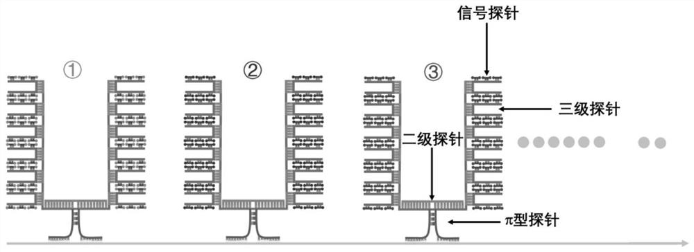
[0060] like figure 1 The single gene detection shown is a method for in situ detection of Actb gene mRNA and DNA in BHK cells by π-FISH. The specific steps are as follows:

[0061] (1) Probe design:

[0062] a. Primary π-type probes: 17 primary π-type probe pairs were designed for the Actb gene in BHK cells, and each primary π-type probe includes a π foot region, a π top region and a π middle region; ,

[0063] The π foot region is the specific hybridization region that is complementary to the base pairing of Actb mRNA, which is represented by NNN… The complementary sequences of the regions are AAGTCCTT and TTCCACTA respectively;

[0064] The sequence on the left side of the primary π-type probe: 5'-NNN...NNNAAGTCCTTNNN...NNN-3'

[0065] The sequence on the right side of the primary π-type probe: 5'-NNN...NNNTTCCACTANNN...NNN-3'

[0066] b. Secondary probe: The secondary probe is divided into a middle region and two end regions; the middle region is a sequence complementa...

Embodiment 2

[0114] like Figures 7 to 8 The single-gene and multi-gene detection in the shown tissue sections, namely, π-FISH in situ detection of Cux2 gene, Pcp4 gene and co-detection of the mRNA of Pcp4 and Rorb genes in mouse brain tissue sections, the specific steps are as follows:

[0115] (1) Probe design:

[0116] a. Primary π-type probes: The design principles of π-type probes for Cux2, Pcp4 and Rorb genes are the same as those in Example 1 a; 19 π-type probes are designed for Cux2 gene, and 8 π-type probes are designed for Pcp4 gene, Rorb gene designed 17 π-type probes, each of which is divided into three regions, namely the π foot region, the π top region and the π middle region. The pi foot region is the specific hybridization region that is complementary to the base pairing of Cux2, Pcp4 and Rorb gene mRNAs, and is represented by NNN... The sequence of the π-type π top region is represented by NNN...

[0117] The sequence information of the primary π-type probe of Cux2 and ...


PUM

PropertyMeasurementUnit
thicknessaaaaaaaaaa

Abstract

The present invention relates to a π-FISH single-molecule probe composition and its application in nucleic acid in situ detection. Type probes, secondary probes, tertiary probes and signal probes; the present invention overcomes traditional fluorescence in situ hybridization, which has low probe specificity, large signal background noise, complex operation, cumbersome steps, and low signal amplification ability and other defects, only a few simple steps of step-by-step amplification probe hybridization can realize the in-situ and single-molecule detection of RNA or DNA nucleic acid information in cells and tissues, which is significantly better than traditional fluorogens in terms of accuracy, throughput and cost. bit hybridization method.

Description

technical field [0001] The invention relates to the technical field of in-situ single-molecule detection of nucleotides, in particular to a π-FISH probe composition for locating single-molecule and its application in nucleic acid in-situ detection. Background technique [0002] In 1969, Gall and Pardue were the first to use radiolabeled probes to interpret nucleic acid information in situ (Pardue 1969), but this method has many shortcomings. First, the radiolabeled probe is a relatively expensive and hazardous material that is unstable in storage and handling; second, the method suffers from high background, long auto-imaging time, and low spatial resolution (Levsky and Singer 2003). Therefore, this method was quickly replaced by fluorescent labeling-based techniques. In 1986, Pinkel et al. used biotin molecules or digoxigenin-conjugated DNA probes, and then used reporter molecules bound to avidin or visualized by anti-digoxigenin antibodies, which improved the detection of...

Claims


Application Information

Patent Timeline
Patent Type & Authority Patents(China)
IPC IPC(8): C12N15/11C12Q1/6841C12Q1/682
CPCC12N15/11C12Q1/6841C12Q1/682C12Q2563/107
Inventor 曹罡戴金霞陶影峰周小六林达徐伟泽
Owner 鲲羽生物科技(江门)有限公司
Learn More