Antigen-antibody co-display method for screening antibody library of membrane antigen
A technology of antibody library and membrane antigen, which is applied in the field of membrane antigen antibody library screening and antigen-antibody co-display, which can solve the problems of inability to perform effective screening, membrane proteins cannot cause sufficient immune response, and antibody display technology cannot be used for library elutriation, etc.
- Summary
- Abstract
- Description
- Claims
- Application Information
AI Technical Summary
Problems solved by technology
Method used
Image

Examples
Embodiment 1
[0115] Example 1. Vector construction and expression localization of multiple transmembrane proteins-antigen proteins
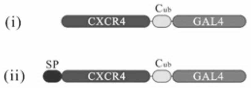
[0116] Purpose: To achieve the effect of localizing in the cell membrane by fused expression of CXCR4 gene with EGFP, Cub, and GAL4 genes.
[0117] Plasmid construction:
[0118] The human CXCR4 (shown in SEQ ID NO.1, specifically:
[0119] MEGISIYTSDNYTEEMGSGDYDSMKEPCFREENANFNKIFLPTIYSIIFLTGIVGNGLVILVMGYQKKLRSMTDKYRLHLSVADLLFVITLPFWAVDAVANWYFGNFLCKAVHVIYTVNLYSSVLILAFISLDRYLAIVHATNSQRPRKLLAEKVVYVGVWIPALLLTIPDFIFANVSEADDRYICDRFYPNDLWVVVFQFQHIMVGLILPGIVILSCYCIIISKLSHSKGHQKRKALKTTVILILAFFACWLPYYIGISIDSFILLEIIKQGCEFENTVHKWISITEALAFFHCCLNPILYAFLGAKFKTSAQHALTSVSRGSSLKILSKGKRGGHSSVSTESESSSFHSS)扩增并测序,与EGFP(SEQ ID NO.2所示,具体为:
[0120] MVSKGEELFTGVVPILVELDGDVNGHKFSVSGEGEGDATYGKLTLKFICTTGKLPVPWPTLVTTLTYGVQCFSRYPDHMKQHDFFKSAMPEGYVQERTIFFKDDGNYKTRAEVKFEGDTLVNRIELKGIDFKEDGNILGHKLEYNYNSHNVYIMADKQKNGIKVNFKIRHNIEDGSVQLADHYQQNTPIGDGPVLLPDNHYLSTQSALSKDPNEKRDHMVLLEFVTAAGITLGMDE...
Embodiment 2
[0127] Example 2. Vector construction and expression localization of antibody modules
[0128] Purpose: To achieve the effect of localization in the cell membrane by fused expression of At1-scFv gene with EGFP, transmembrane peptide TMpho, and NubG.
[0129] Plasmid construction:
[0130] The anti-human CXCR4 single-chain antibody At1-scFv (shown in SEQ ID NO.6, specifically:
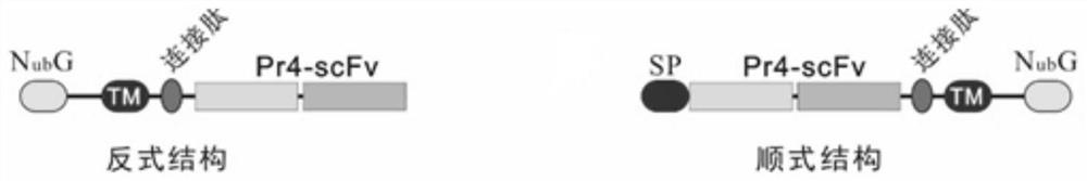
[0131] DIVMTQSPDSLAVSLGERATINCKSSQSLFNSRTRKKYLAWYQQKPGQPPKLLIYWASKRKSGVPDRFSGSGSGTDFTLTISSLQAEDVAVYYCKQSRFLRAFGQGTKLEIKGGGGSGGGGSGGGGSEVQLVESGGGLVQPGGSLRLSCAASGFTSTDYYFSWVRQAPGKGLEWVGFIRTKSKGYTTEYSGSVKGRFTISRDDSKNSLYLQMNSLKTEDTAVYYCAREPITTDPRDYWGQGTLVTVS)编码序列扩增并测序,与EGFP、泛素蛋白N端结构域NubG(SEQ ID NO.7所示,具体为:MQIFVKTLTGKTGTLEVESSDTIDNVKSKIQDKEGIP)、酵母跨膜肽TMpho(SEQ ID NO.8所示,具体 is: SWLLAFAGCKPRNVLLMAMCVVFFLSMWISNVAAPVLTYSLLSP), these four parts are fused, and the whole fusion protein is cloned into the yeast expression vector pGAD-T7 to form as Figure 8 The structure shown in A is designated as pGBK-T7-NubG-TMph...
Embodiment 3
[0136] Example 3. Interaction detection of CXCR4 single chain antibody At1-scFv with CXCR4 antigen
[0137] Objective: To observe the interaction between CXCR4 and its single-chain antibody At1-scFv.
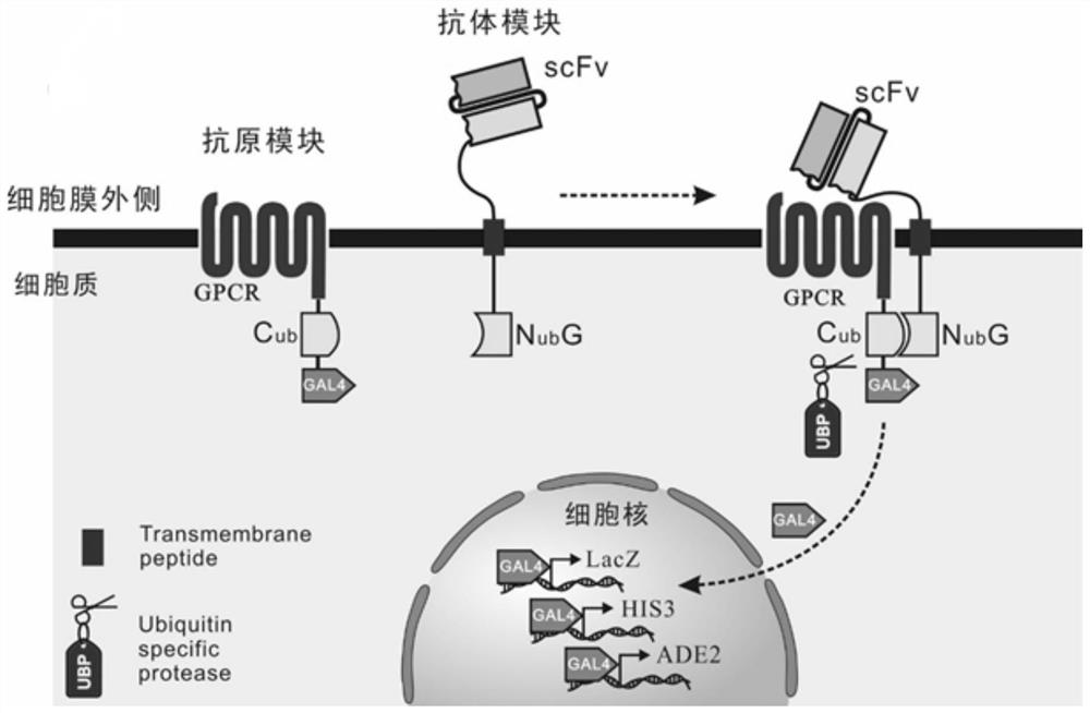
[0138] Principle: When the CXCR4 protein interacts with the antibody, the intracellular ubiquitin Cub and Nub domains are pulled together to form an active and complete ubiquitin protein, which is then digested by the endogenous ubiquitin-specific protease of the cell to release the Cub The transcription factor GAL4 connected at the end enters the nucleus to transcribe the reporter genes His3, Ade2 and LacZ. The first two reporter genes help cells grow on SD-His-Ade-auxotrophic medium, and the latter catalyzes the development of X-a-gal substrate in the medium. Among them, antibody plasmids have two topological forms, namely Cis and Trans.
[0139] Plasmid construction:
[0140] Antigen plasmid: Human CXCR4 gene, Cub and GAL4 gene were fused to pGAD-T7 plasmid, which was desi...
PUM
 Abstract
Description
Claims
Application Information
 - Generate Ideas
- Intellectual Property
- Life Sciences
- Materials
- Tech Scout
- Unparalleled Data Quality
- Higher Quality Content
- 60% Fewer Hallucinations
Browse by: Latest US Patents, China's latest patents, Technical Efficacy Thesaurus, Application Domain, Technology Topic, Popular Technical Reports.
© 2025 PatSnap. All rights reserved.Legal|Privacy policy|Modern Slavery Act Transparency Statement|Sitemap|About US| Contact US: help@patsnap.com