
ACE2-Fc fusion protein function test method for treating COVID-19

A COVID-19, fusion protein technology, applied in the field of functional testing of ACE2-Fc fusion protein for the treatment of COVID-19

Pending Publication Date: 2021-01-15
苏州新格诺康生物技术有限公司
View PDF0 Cites 9 Cited by
  • Summary
  • Abstract
  • Description
  • Claims
  • Application Information

AI Technical Summary

Problems solved by technology

While neutralizing antibodies are a very attractive option, developing humanized antibodies against the S protein or ACE2 with sufficient affinity and specificity to neutralize the virus may take months or longer

Method used


Image

  • ACE2-Fc fusion protein function test method for treating COVID-19
  • ACE2-Fc fusion protein function test method for treating COVID-19
  • ACE2-Fc fusion protein function test method for treating COVID-19

Examples


Embodiment

[0030] A method for testing the function of an ACE2-Fc fusion protein for treating COVID-19, specifically comprising the following:

[0031] Step 1. Expression of human wild-type ACE2-Fc fusion protein:

[0032] (1) Cloning the DNA sequence of the extracellular domain (ECD) of human wild-type ACE;

[0033] (2) Fusing the ECD of ACE2 with the Fc part of human IgG1 as follows figure 1 Prolong the half-life of soluble ACE2;

[0034] (3) expressing the ACE2-Fc fusion protein in CHO cells, and using protein A / G agarose to purify the ACE2-Fc fusion protein in the cell culture supernatant;

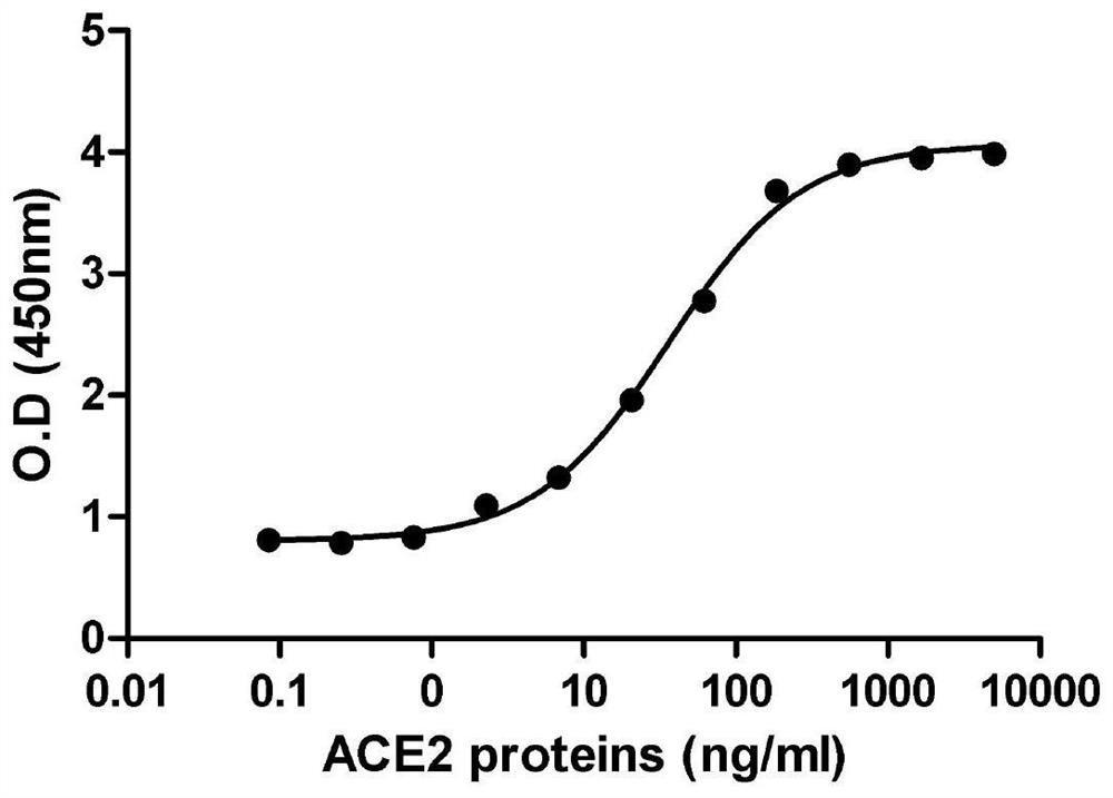
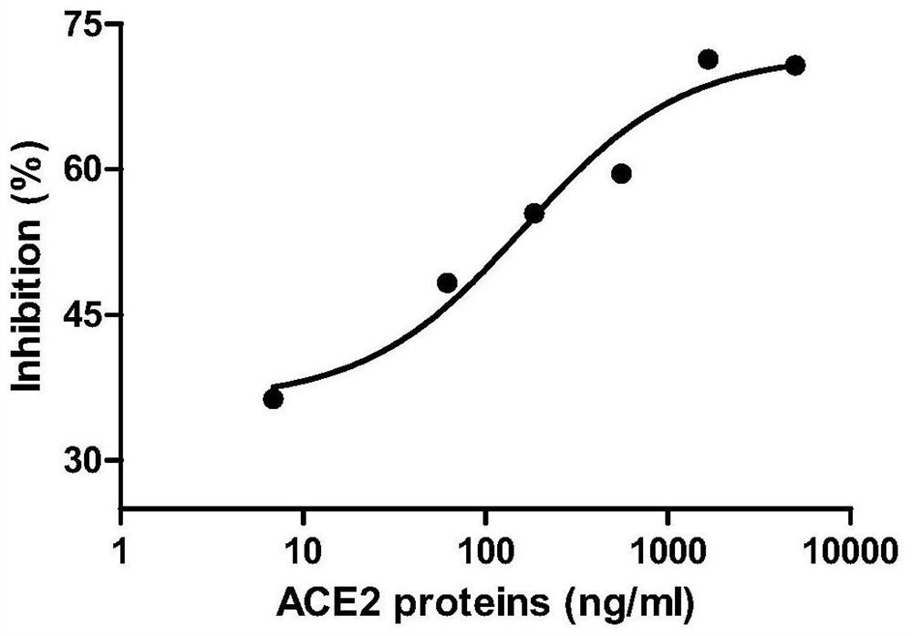
[0035] Step 2. In vitro testing of the binding function and inhibitory function of the ACE2-Fc fusion protein to the SARS-CoV-2 S1 / RBD protein:

[0036] (4) Coat a 96-well plate with 10ug / ml of SARS-CoV-2RBD-His fusion protein, and then use ACE2-Fc fusion protein to titrate with 2-fold serial dilutions starting from 5ug / ml;

[0037] (5) Record the test data obtained from the titration experim...


PUM


Abstract

The invention provides an ACE2-Fcfusion protein function test method for treating COVEID-19. The ACE2-Fc fusion protein function test method specifically comprises the following steps of expressing ahuman wild type ACE2-Fc fusion protein: cloning a DNA sequence of an extracellular domain (ECD) of human wild type ACE; enabling the ECD of the ACE2 to be fused with the Fc part of the human IgG1, sothat the half-life period of the soluble ACE2 is prolonged; expressing the ACE2-Fc fusion protein in CHO cells, and purifying the ACE2-Fc fusion protein in a cell culture supernatant by using proteinA / G agarose; and optimized an expression system. The invention effectively proves that the ACE2-Fc fusion protein can prevent or block viruses from entering cells, slow down the infection process andexpose the viruses to the outside of the cells so as to be recognized and eliminated by a human immune system.

Description

technical field [0001] The invention relates to the technical field of fusion proteins, in particular to a method for testing the function of ACE2-Fc fusion proteins for treating COVID-19. Background technique [0002] The coronavirus first binds to the target receptor on the surface of the host cell through protein spikes, and after fusion with the cell membrane, the viral nucleocapsid enters the cell for subsequent replication. In SARS-CoV cases, the spike protein on the surface of the virion mediates receptor recognition and membrane fusion. During viral infection, the trimeric S protein is cleaved into S1 and S2 subunits, and the S1 subunit is released during the transition to the postfusion conformation. S1 contains the receptor-binding domain (RBD), which directly binds to the peptidase domain (PD) of ACE2, while S2 is responsible for membrane fusion. Given the high sequence homology (70–80%) between SARS-CoV-2 and SARS-CoV, it has recently been shown that the extrac...

Claims


Application Information

Patent Timeline
Patent Type & Authority Applications(China)
IPC IPC(8): C12N9/48G01N33/569G01N33/573G01N33/68
CPCC12N9/485G01N33/56983G01N33/573G01N33/6893C12Y304/17023C07K2319/30G01N2333/165G01N2333/948
Inventor 谢志伟杨小丽吴畏白彩虹严俊
Owner 苏州新格诺康生物技术有限公司
Learn More