External suction tube of gastroscope

A suction tube, external technology, applied in the directions of gastroscope, endoscope, esophagoscope, etc., can solve the problems of difficult control of suction tube, difficult pylorus, and high difficulty in operation

Pending Publication Date: 2021-01-05
汪剑波
View PDF0 Cites 0 Cited by
  • Summary
  • Abstract
  • Description
  • Claims
  • Application Information

AI Technical Summary

Problems solved by technology

[0006] 1. Turn over and observe the patient on the left side, and then turn the patient on the right side to expose the remaining gastric fundus mucosa; however, patients with acute upper gastrointestinal bleeding often need to complete the treatment under tracheal intubation, and it is difficult and troublesome to turn over first , and the degree is limited, so part of the gastric fundus mucosa still cannot be fully exposed
[0007] 2. Send the blood clot into the intestinal tract with a snare; this scheme is very difficult to operate. It is already very difficult to enter the pylorus with a transparent cap, and it is even more difficult to enter the descending part of the duodenum with a snare with a blood clot , and it is also very inconvenient to separate the blood clot in the descending part. This operation cannot be completed at one time, and repeated operations are required many times
[0008] 3. Use forceful suction or use a snare to remove the blood clot in batches; this plan also requires repeated operations, which is very troublesome. If the bleeding focus under the blood clot is still actively bleeding, the speed of removal may be slower than Not on the speed of bleeding
[0009] 4. Send an external suction tube to suck out blood clots, etc.; this scheme is feasible, but it is difficult to send a large suction tube into the gastric cavity, and a small suction tube cannot effectively attract blood clots, and there are also disadvantages It is difficult to control the suction tube in the gastric cavity, and it is impossible to quickly remove the blood clot. At the same time, because of the strong suction, it is easy to suck the gastric mucosa firmly, which hinders the suction of the blood clot and damages the normal mucosa
The results show that there is currently no clinically effective method for removing blood clots in the fundus of the stomach under early endoscopy

Method used


Image

  • External suction tube of gastroscope
  • External suction tube of gastroscope

Examples


Embodiment 1

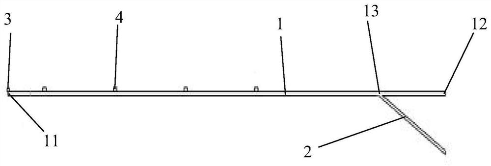
[0033] see figure 1 , this embodiment provides a gastroscope external suction tube, including: suction tube 1, auxiliary tube 2, connecting part-3;

[0034] The head end of the suction tube is provided with an air-introducing port 12, the end is provided with a suction port 11, and the middle and upper part is provided with a through-port 13;

[0035] The auxiliary pipe 2 is connected through the through opening 13 to the suction pipe 1 , and the connection part 1 is connected to the suction port 11 in a dislocation manner.

[0036] In this embodiment, the connecting part one 3 is a transparent cap for connecting the endoscope, and the dislocation connection can prevent the suction tube from sucking the gastric mucosa firmly.

[0037] In one embodiment, the inner diameter of the suction tube 1 is 5 mm, the outer diameter is 7 mm, and the length is 130 cm.

[0038] In one embodiment, the dislocation distance of the dislocation connection is 3mm.

[0039] In one embodiment, t...

Embodiment 2

[0043] This embodiment provides an external suction tube for a gastroscope, including: a suction tube 1, an auxiliary tube 2, and a connecting part-3;

[0044] The head end of the suction tube is provided with an air-introducing port 12, the end is provided with a suction port 11, and the middle and upper part is provided with a through-port 13;

[0045] The auxiliary pipe 2 is connected through the through opening 13 to the suction pipe 1 , and the connection part 1 is connected to the suction port 11 in a dislocation manner.

[0046] In the second embodiment, the first connecting part 3 is a transparent cap for connecting the endoscope, and the dislocation connection can prevent the suction tube from sucking the gastric mucosa firmly.

[0047] In the second embodiment, the inner diameter of the suction tube 1 is 5 mm, the outer diameter is 7 mm, and the length is 125 cm.

[0048] In the second embodiment, the dislocation distance of the dislocation connection is 3 mm.

[0...

Embodiment 3

[0053] This embodiment provides an external suction tube for a gastroscope, including: a suction tube 1, an auxiliary tube 2, and a connecting part-3;

[0054] The head end of the suction tube is provided with an air-introducing port 12, the end is provided with a suction port 11, and the middle and upper part is provided with a through-port 13;

[0055] The auxiliary pipe 2 is connected through the through opening 13 to the suction pipe 1 , and the connection part 1 is connected to the suction port 11 in a dislocation manner.

[0056] In the third embodiment, the connecting part one 3 is a transparent cap for connecting the endoscope, and the dislocation connection can prevent the suction tube from sucking the gastric mucosa firmly.

[0057] In the third embodiment, the inner diameter of the suction tube 1 is 5 mm, the outer diameter is 7 mm, and the length is 135 cm.

[0058] In the third embodiment, the dislocation distance of the dislocation connection is 4 mm.

[0059] In...


PUM


Abstract

The invention discloses an external suction tube of a gastroscope. The external suction tube comprises a suction tube body, an auxiliary tube and a first connecting part, wherein the suction tube bodycomprises a suction port and an air guide port which are formed in the two ends and a through port which is formed in the middle and is close to the air guide port, the auxiliary tube body is connected with the suction tube body in a through mode through the through port, and the first connecting part is connected with the suction port in a staggered manner; The external suction tube further comprises a plurality of second connecting parts fixedly connected with the suction tube; and the suction tube, the auxiliary tube and the first connecting part are all made of rubber. The suction tube ismoderate in diameter, can easily enter a gastric cavity along with the gastroscope, can freely control the angle through the gastroscope and cannot firmly suck gastric mucosa, the gastroscope does not need to withdraw after blood clots are treated, and related hemostatic treatment can be continued.

Description

technical field [0001] The invention relates to the technical field of medical instruments, in particular to a gastroscope external suction tube. Background technique [0002] Acute upper gastrointestinal bleeding is one of the common critical illnesses in the Department of Gastroenterology. It often presents with hematemesis and melena as the main clinical manifestations. The condition is dangerous and the mortality rate is high. According to the etiology, it can be divided into non-variceal bleeding and variceal bleeding . In recent years, with the continuous development of treatment technology, its incidence has shown a significant downward trend, but its related mortality rate is still as high as 2-10%, and about 33% of patients will have recurrent upper gastrointestinal bleeding. growing public health problem. Early endoscopic examination and treatment can significantly reduce the mortality rate and rebleeding rate of upper gastrointestinal bleeding. Therefore, early...

Claims


Application Information

Patent Timeline
Patent Type & Authority Applications(China)
IPC IPC(8): A61B1/012A61B1/015A61B1/273
CPCA61B1/012A61B1/015A61B1/2736
Inventor 汪剑波
Owner 汪剑波
Why Eureka
Try Eureka