
M protein ultraviolet spectroscopy detection method

A spectroscopic detection and ultraviolet technology, applied in the field of M protein detection, can solve the problems of not being able to directly prompt whether patients have M protein, expensive equipment and reagent consumables, cumbersome operation steps, etc. Interpreting Simple Effects

Pending Publication Date: 2020-12-29
范庆坤
View PDF0 Cites 0 Cited by
  • Summary
  • Abstract
  • Description
  • Claims
  • Application Information

AI Technical Summary

Problems solved by technology

These two methods are manual or semi-automatic. The operation steps are cumbersome, and the instruments and reagent consumables are expensive. Each time, batch testing is required, and the amount of testing is small, and the testing takes a long time. Only a few hospitals in the country can perform testing, and it is difficult to popularize. To all testing laboratories across the country
Quantitative detection of total protein, albumin and hemoglobin is also commonly used as an indication for early screening of M proteinemia, but not all patients have been tested clinically
Therefore, in daily clinical diagnosis and treatment, the missed diagnosis rate of M proteinemia is as high as 60%. On the one hand, it is due to the lack of understanding of M proteinemia by clinicians, and on the other hand, it is mainly because routine testing items cannot directly indicate whether patients have M protein
At present, there is no conventional biochemical detection reagent or detection equipment on the market that can detect M protein

Method used


Image

  • M protein ultraviolet spectroscopy detection method

Examples


Embodiment Construction

[0033] The specific implementation manners of the present invention will be described in detail below in conjunction with the accompanying drawings. The described embodiments are only illustrations and explanations of the present invention, and do not constitute the only limitation of the present invention.

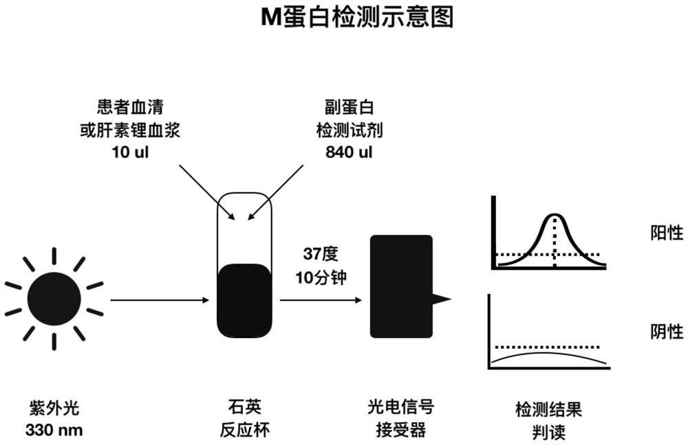
[0034] Such as figure 1 Shown, the inventive method comprises steps:

[0035] a) Take 1 ml of the subject's venous blood and place it in a vacuum lithium heparin anticoagulation test tube or a vacuum additive-free test tube or other test tubes;

[0036] b) If a test tube without additives is used, it needs to be left for at least 10 minutes, and the sample pretreatment is performed after the blood is naturally coagulated; if it is a heparin lithium test tube, the sample pretreatment can be performed directly;

[0037] c) Sample pretreatment requirements: centrifuge the above-mentioned completely coagulated blood test tubes or heparin lithium anticoagulated blood test tub...


PUM


Abstract

The invention discloses an M protein ultraviolet spectroscopy detection method, and belongs to the technical field of M protein detection. The detection method is implemented through the reaction ratio of the patient sample size to the accessory protein detection reagent dosage and by combining a light source, operation is easy and convenient, result interpretation is easy, and the positive resultspecificity is high. The method is suitable for all kinds of serum or plasma and is not affected by anticoagulants. Detection can be carried out as long as venous blood or arterial blood sampling iscarried out on patients. The method is low in detection reagent cost, all medical laboratories can bear the method at present, and the problem that the patients are hard to see a doctor and expensivecan be relieved to a great extent. According to the method, large-scale general screening is carried out on all the patients in the medical laboratories, the clinical laboratories are promoted to findsuspicious M proteinemia patients in the early stage, and the patients can be diagnosed and treated early.

Description

technical field [0001] The invention belongs to the technical field of M protein detection, and in particular relates to an M protein ultraviolet spectroscopic detection method. Background technique [0002] Multiple myeloma is a malignant disease of abnormal proliferation of clonal plasma cells, and it is the second most common malignant tumor of the blood system in many countries. It mostly occurs in the elderly and cannot be completely cured at present, but early diagnosis, regular monitoring, and early intervention can prolong the survival time and improve the quality of life of patients. The detection of M protein is the core means of early diagnosis of multiple myeloma. M protein is an abnormal immunoglobulin or immunoglobulin fragment in human blood, which is mainly produced by the malignant proliferation of plasma cells or B lymphocytes. Clinical often called monoclonal protein, monoclonal immunoglobulin or myeloma protein. In addition to multiple myeloma (Multipl...

Claims


Application Information

Patent Timeline
IPC IPC(8): G01N21/33
CPCG01N21/33
Inventor 范庆坤
Owner 范庆坤
Learn More