
Method for in-situ preparation of TEM sample of nanoscale particles in sample with complex structure

A complex structure, in-situ preparation technology, applied in the fields of planetary science and planetary exploration, can solve the problems of wasting sample resources, unavoidable sample contamination, inability to perform polishing, etc., to reduce pollution, simplify operation procedures, and overcome area damage. Effect

Active Publication Date: 2020-09-22
INST OF GEOCHEM CHINESE ACADEMY OF SCI +1
View PDF5 Cites 1 Cited by
  • Summary
  • Abstract
  • Description
  • Claims
  • Application Information

AI Technical Summary

Problems solved by technology

[0005] In view of the waste of sample resources in the preparation of TEM flake samples by the existing conventional FIB technology, the inability to avoid sample contamination during processing, and the inability to apply to nano-scale particle samples, the present invention provides a method for in-situ preparation of nano-scale particles in samples with complex structures. The method of TEM samples to realize the preparation of TEM samples of nanoscale particles entrapped in bulk samples with complex structures and unable to be polished or scarce extraterrestrial micron-scale powder samples

Method used


Image

  • Method for in-situ preparation of TEM sample of nanoscale particles in sample with complex structure
  • Method for in-situ preparation of TEM sample of nanoscale particles in sample with complex structure
  • Method for in-situ preparation of TEM sample of nanoscale particles in sample with complex structure

Examples


Embodiment 1

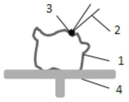
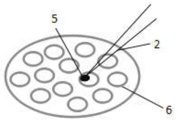
[0036] In this example, a TEM sample of nanoscale particles in samples with a complex structure was prepared in situ using a double beam electron microscope, and a schematic diagram of extracting nanoscale particles on the sample surface and fixing nanoscale particles on a copper grid by using a double beam electron microscope is as follows Figure 1~2 As shown, the specific steps are as follows:

[0037] (1) The sample used in this example is a dolomite block sample, which has irregular shape, uneven appearance, complex surface phase structure, and differences in composition and morphology of different parts of the surface, so the surface of the sample cannot be pre-polished. And there are nanoscale particles on the sample, and these nanoscale particles are aggregated together to form nanoparticle clusters.

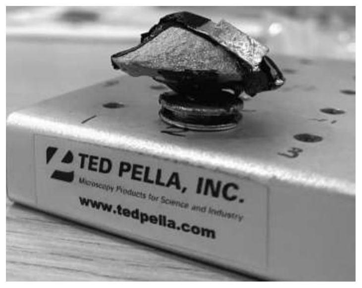
[0038] Fix the sample on the sample holder with carbon conductive tape and copper conductive tape, such as image 3 As shown, the sample holder is then placed on the sa...

Embodiment 2

[0043] In this example, a TEM sample of nanoscale particles in a sample with a complex structure was prepared in situ using a double-beam electron microscope, and the steps were as follows:

[0044] (1) The sample used in this example is a simulated lunar soil sample. In order to ensure in-situ extraction, the sample is not suitable for dispersion treatment with alcohol, and there are nano-scale particles on the sample, and these nano-scale particles gather together to form nano-particle clusters cluster.

[0045] Fix the sample on the sample holder with carbon conductive tape, then place the sample holder on the sample stage in the sample chamber of the double-beam electron microscope, install the copper mesh in the slot horizontal to the sample stage, and make the copper mesh and the sample stage On the same level, seal the sample chamber and evacuate the sample chamber. After the vacuum degree of the sample chamber meets the requirements, use the scanning electron microscop...

Embodiment 3

[0050] In this example, a TEM sample of nanoscale particles in a sample with a complex structure was prepared in situ using a double-beam electron microscope, and the steps were as follows:

[0051] (1) The sample micron SiO used in this embodiment 2 A powder sample that has nanoscale particles on its surface that aggregate together to form nanoparticle clusters.

[0052] Fix the sample on the sample holder, then place the sample holder on the sample stage in the sample cavity of the double-beam electron microscope, install the copper mesh in the slot horizontal to the sample stage, so that the copper mesh and the sample stage are on the same level , seal the sample cavity and evacuate the sample cavity. After the vacuum degree of the sample cavity meets the requirements, use the scanning electron microscope function of the double-beam electron microscope to observe the surface morphology of the sample and find the nanoparticle cluster area of ​​interest, such as Image 6 As ...


PUM

PropertyMeasurementUnit
Diameteraaaaaaaaaa

Abstract

The invention provides a method for in-situ preparation of a TEM sample of nanoscale particles in a sample with a complex structure. The method comprises steps of placing a sample on a sample table ina sample cavity of a double-beam electron microscope, arranging a copper net on the sample table, sealing the sample cavity and vacuumizing, observing the surface morphology of the sample by using ascanning electron microscope function, finding out an interested nano particle cluster region, keeping the sample table in a horizontal state, adsorbing the nanoscale particles to a needle tip of thenano manipulator by virtue of electrostatic adsorption force, retracting the nano manipulator, moving the copper net to a position right below the electronic gun, fixing nanoscale particles adsorbed on a needle tip to an edge of a copper hole in the copper net by virtue of the electrostatic adsorption force, and taking out the copper net fixed with the nanoscale particles so as to finish sample preparation. The method is advantaged in that the preparation of the TEM sample of nano-scale particles entrained in a blocky sample or a rare extraterrestrial micron-scale powder sample which has a complex structure and cannot be subjected to polishing treatment can be realized, and the method can be applied to the basic research field of planetary science and planetary detection.

Description

technical field [0001] The invention belongs to the field of planetary science and planetary exploration, and relates to a method for in-situ preparation of a TEM sample of nano-scale particles by using a double-beam electron microscope, more specifically, a method for in-situ preparation of a TEM sample of nano-scale particles in samples with complex structures. Background technique [0002] In the past ten years, the nanoprocessing technology of focused ion beam-electron beam (FIB / SEM) electron microscope (dual-beam electron microscope), as an important tool for transmission electron microscope (TEM) sample preparation, has been widely used in materials, biology, and geochemistry. , planetary science, and geology. Compared with the process of preparing TEM samples by ultramicrotome and ion thinner, the biggest advantage of using double-beam electron microscope is that it can prepare TEM samples in situ. With the demand for in-depth exploration and development of the micro...

Claims


Application Information

Patent Timeline
IPC IPC(8): G01N23/2005
CPCG01N23/2005
Inventor 李瑞李阳钟怡江李雄耀刘建忠王世杰
Owner INST OF GEOCHEM CHINESE ACADEMY OF SCI
Learn More