
Hip joint image processing method and system based on magnetic resonance, storage medium and equipment

A magnetic resonance image and image processing technology, applied in image data processing, image enhancement, image analysis, etc., can solve the problems of classification, inability to objectively describe the value of morphological parameters, and the influence of human factors.

Active Publication Date: 2020-08-21
SUZHOU INST OF BIOMEDICAL ENG & TECH CHINESE ACADEMY OF SCI
View PDF1 Cites 6 Cited by
  • Summary
  • Abstract
  • Description
  • Claims
  • Application Information

AI Technical Summary

Problems solved by technology

In addition, clinicians often only pay attention to the changes of one or several morphological parameters of the hip joint in children with DDH, without detailed classification and comprehensive observation of various morphological changes, which makes DDH have a certain probability of misdiagnosis
However, there is no such a set of methods and systems for systematically judging the degree of disease in children with DDH combined with magnetic resonance images of children with DDH.
[0004] In addition, in clinical diagnosis and treatment, the observation of the morphological parameters of the hip joint remains at naked eye observation or manual measurement, which requires high professionalism of doctors
Moreover, the manual measurement process makes it time-consuming and labor-intensive to judge whether the morphological parameters are normal from the magnetic resonance images, and the manual measurement results are greatly affected by human factors, which cannot objectively describe the values ​​of the morphological parameters.
However, there is no such a processing method that comprehensively measures the morphological parameters of the hip joint.

Method used


Image

  • Hip joint image processing method and system based on magnetic resonance, storage medium and equipment
  • Hip joint image processing method and system based on magnetic resonance, storage medium and equipment
  • Hip joint image processing method and system based on magnetic resonance, storage medium and equipment

Examples


Embodiment Construction

[0084] Below, the present invention will be further described in conjunction with the accompanying drawings and specific implementation methods. It should be noted that, under the premise of not conflicting, the various embodiments described below or the technical features can be combined arbitrarily to form new embodiments. .

[0085] The present invention provides a hip joint image processing method and system based on magnetic resonance, which provides comprehensive and reliable support for the diagnosis and treatment of clinical DDH. All test tasks can be written and stored in script files, and can also be simulated line by line at the system terminal Execution; In addition, the system can work in offline mode, that is, run on a computer or workstation, and can also be deployed on terminal electronic devices, such as mobile phones or tablets, to achieve online programmable design. After the calculation is completed, the simulation data results can be transmitted after the ...


PUM


Abstract

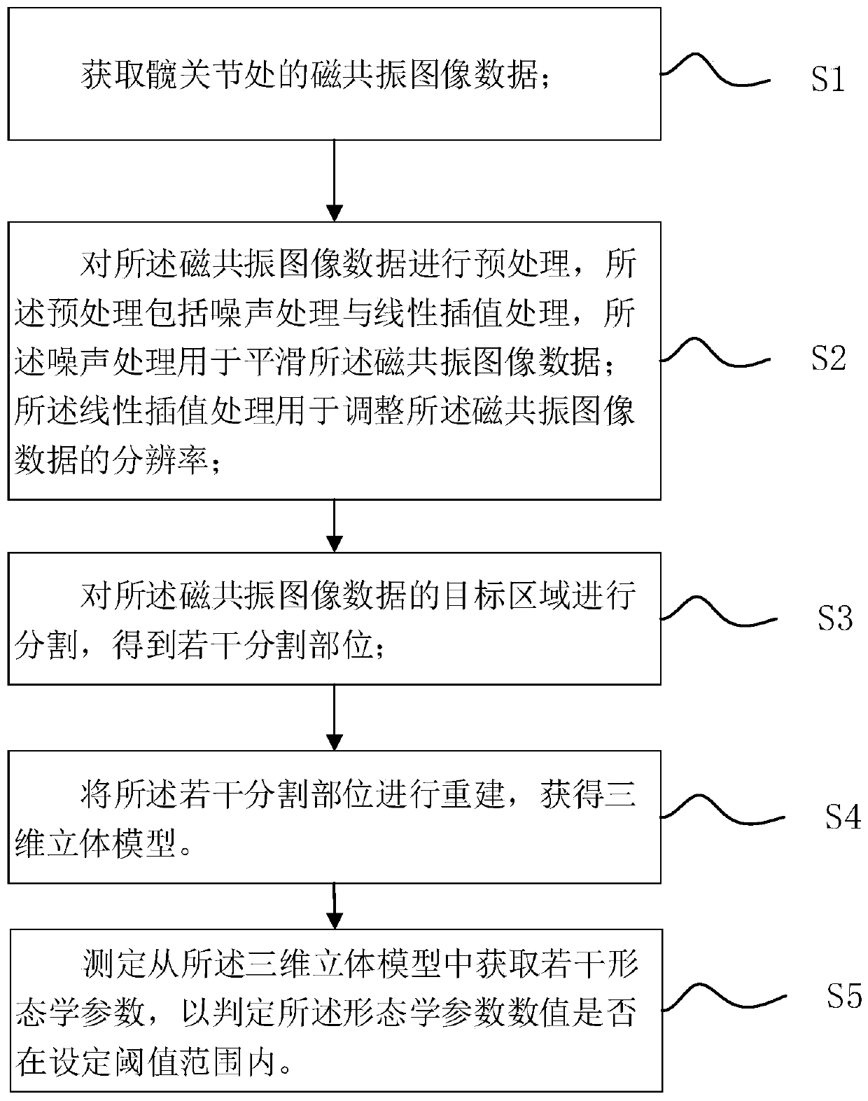
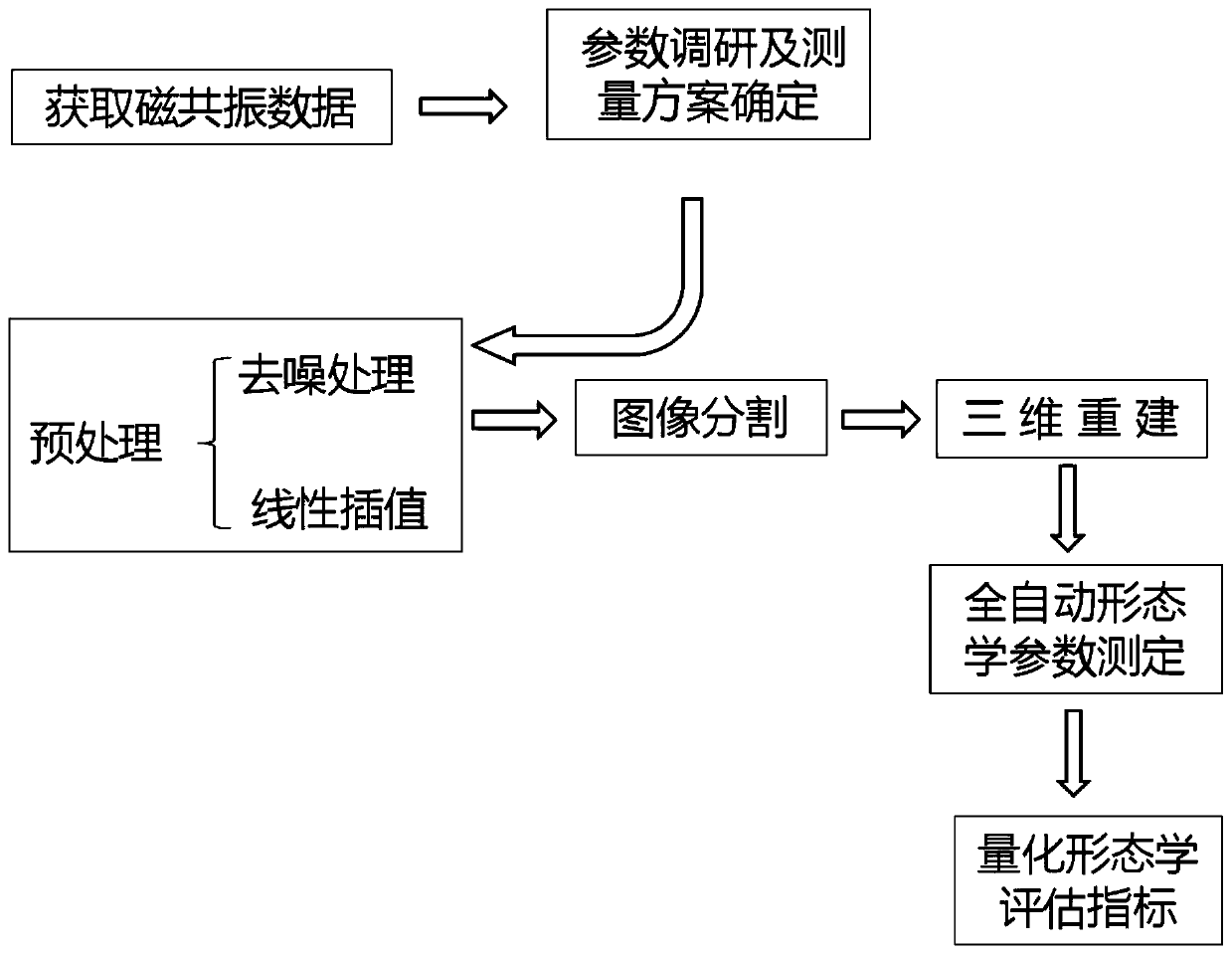
The invention provides a hip joint image processing method based on magnetic resonance. The method comprises the following steps: acquiring magnetic resonance image data at a hip joint; segmenting a target area of the magnetic resonance image data to obtain a plurality of segmented parts; reconstructing the plurality of segmented parts to obtain a three-dimensional model; and measuring a pluralityof morphological parameters obtained from the three-dimensional model to determine whether the numerical values of the morphological parameters are within a set threshold range or not. The inventionfurther provides a hip joint image processing system based on magnetic resonance, a storage medium and equipment. According to the invention, the hip joint development condition can be accurately measured; the method not only can provide a basis for disease diagnosis and preoperative planning, but also can provide convenience for postoperative long-term dynamic monitoring, is a whole-process quantitative evaluation means capable of penetrating through early diagnosis, treatment and postoperative dynamic monitoring of DDH diseases, and is of great significance to precision medical treatment ofthe diseases and healthy growth of sick children.

Description

technical field [0001] The invention relates to the field of magnetic resonance quantitative systems, in particular to a magnetic resonance-based hip joint image processing method. Background technique [0002] Developmental dysplasia of the hip (DDH), also known as developmental dysplasia of the hip, is the most common hip disease in pediatric orthopedics. As the age of the children increases, the lesion becomes more complicated, the treatment becomes more difficult, and the curative effect becomes worse. Therefore, early diagnosis and treatment of DDH are very important. After DDH surgery, some children have residual deformities of varying degrees. Therefore, children with DDH need to be followed up for a long time until the skeletal development stops in adolescence, and the problems discovered during the follow-up process should be dealt with in time. Research and development of imaging quantitative evaluation methods for developmental dislocation of the hip, to achieve ...

Claims


Application Information

Patent Timeline
IPC IPC(8): G06T7/11G06T7/155G16H30/20G06T5/00
CPCG06T7/11G06T7/155G16H30/20G06T2207/30008G06T5/70Y02A90/30
Inventor 吴明娣杨晓冬盛茂冯洁贾慧惠吴继志张欣常严
Owner SUZHOU INST OF BIOMEDICAL ENG & TECH CHINESE ACADEMY OF SCI
Learn More