
Automatic liver segmentation method based on deformation model of CT image

A deformation model and CT image technology, applied in the field of image processing, can solve problems such as large registration errors, inability to realize automatic liver segmentation batch processing, and impact on segmentation results, and achieve strong robustness

Inactive Publication Date: 2020-07-28
HARBIN UNIV OF SCI & TECH
View PDF2 Cites 0 Cited by
  • Summary
  • Abstract
  • Description
  • Claims
  • Application Information

AI Technical Summary

Problems solved by technology

[0002] At present, the liver segmentation method is generally based on the active shape model (ASM) liver semi-automatic segmentation method, although these methods can effectively segment the liver tissue, but in the initialization stage of the liver shape, manual control and interaction are required. Artificially manually select a seed point at the main branch of the portal vein and the hepatic vein to roughly segment the liver vessels, and manually cut off the leaking adjacent organs at the entrance of the portal vein and the hepatic vein
This makes the whole method unable to achieve automatic liver segmentation batch processing, and a major shortcoming of the semi-automatic segmentation method is that the segmentation results will be affected by human factors, which in turn affects the repeatability of the segmentation results
The existing atlas-based segmentation methods still have the following two main disadvantages: (1) the constructed liver atlas tends to be biased towards the specific anatomical structure of the selected initial template image; (2) it is easy to align the grayscale image of the liver atlas to the target image. Produces large registration errors, especially in segmenting liver cases adjacent to organs with similar gray values ​​and containing large lesions

Method used


Image

  • Automatic liver segmentation method based on deformation model of CT image
  • Automatic liver segmentation method based on deformation model of CT image
  • Automatic liver segmentation method based on deformation model of CT image

Examples


Embodiment Construction

[0023] The following will clearly and completely describe the technical solutions in the embodiments of the present invention with reference to the accompanying drawings in the embodiments of the present invention. Obviously, the described embodiments are only some, not all, embodiments of the present invention. Based on the embodiments of the present invention, all other embodiments obtained by persons of ordinary skill in the art without making creative efforts belong to the protection scope of the present invention.

[0024] In order to make the above objects, features and advantages of the present invention more comprehensible, the present invention will be further described in detail below in conjunction with the accompanying drawings and specific embodiments.

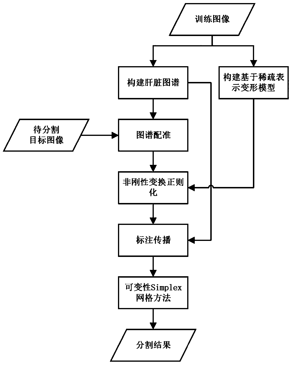
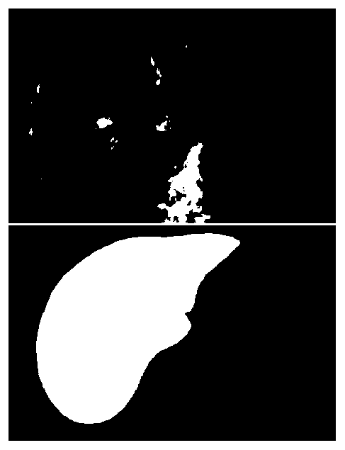
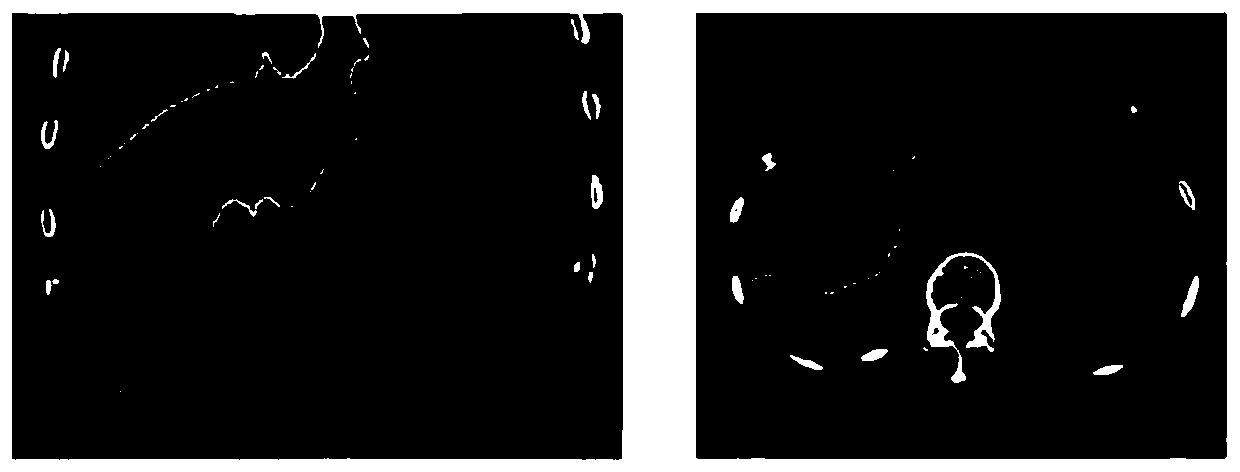
[0025] refer to Figure 1-3 , the present invention provides an automatic liver segmentation method based on a deformation model of CT images. In the training phase, two models that need to be used in the atlas-ba...


PUM


Abstract

The invention discloses an automatic liver segmentation method based on a deformation model of a CT image. The method comprises the steps: 1, building a liver atlas, and representing the deformation model SRDM based on sparsity, and the liver atlas comprises a gray level image and a marking image corresponding to the gray level image; 2, performing liver map registration on a to-be-segmented target image, and constructing a non-rigid transformation model for aligning a grayscale image of the liver map to the target image; 3, regularizing the non-rigid transformation model in the step 2 by using a sparse representation deformation model SRDM; 4, propagating the labeled image of the liver map to a target image by using the regularized transformation model to obtain an initial segmentation result; and step 5, for the data with relatively large segmentation errors, carrying out fine segmentation on an initial segmentation result. Through the scheme, the segmentation precision close to thatof a semi-automatic segmentation method is obtained, and the experimental result can be repeated.

Description

technical field [0001] The invention relates to the technical field of image processing, in particular to an automatic liver segmentation method based on a deformation model of a CT image. Background technique [0002] At present, the liver segmentation method is generally based on the active shape model (ASM) liver semi-automatic segmentation method, although these methods can effectively segment the liver tissue, but in the initialization stage of the liver shape, manual control and interaction are required. Artificially manually select a seed point at the main branch of the portal vein and the hepatic vein to roughly segment the liver vessels, and manually cut off the leaking adjacent organs at the entrance of the portal vein and the hepatic vein. This makes the whole method unable to achieve automatic liver segmentation batch processing, and a major deficiency of the semi-automatic segmentation method is that the segmentation results will be affected by human factors, wh...

Claims


Application Information

Patent Timeline
IPC IPC(8): G06T7/136G06T7/33
CPCG06T2207/10081G06T2207/20081G06T2207/30056G06T2207/30204G06T7/136G06T7/344
Inventor 王进科耿晓旭郭广寒侯甲童李响
Owner HARBIN UNIV OF SCI & TECH
Learn More