Multi-detector X-ray fluorescence micro-area scanning instrument and imaging method thereof

A technology of scanning instruments and imaging methods, which is applied in the direction of instruments, scientific instruments, measuring devices, etc., can solve the problems of immature manufacturing technology, astonishing dead time, and low luminous flux of X-fluorescence signals, achieve high-precision and high-efficiency scanning, increase Effects of detection area and count rate and improvement of scanning efficiency

Pending Publication Date: 2020-07-28
NCS TESTING TECH
View PDF1 Cites 1 Cited by
  • Summary
  • Abstract
  • Description
  • Claims
  • Application Information

AI Technical Summary

Problems solved by technology

[0005] The above existing technical solutions have the following defects: due to the low generation efficiency of X-rays and the lower luminous flux of X-fluorescence signals, only when the detected fluorescence counts reach a certain number in actual detection can a relatively high-precision signal be obtained. Test results
At present, the manufacturing technology of a single large-area high-resolution X-ray energy spectrum detector on the market is still immature, and the dead time caused by high-throughput X-ray processing will be very alarming, and the resolution and other indicators cannot meet the requirements.

Method used


Image

  • Multi-detector X-ray fluorescence micro-area scanning instrument and imaging method thereof
  • Multi-detector X-ray fluorescence micro-area scanning instrument and imaging method thereof
  • Multi-detector X-ray fluorescence micro-area scanning instrument and imaging method thereof

Examples


Embodiment 1

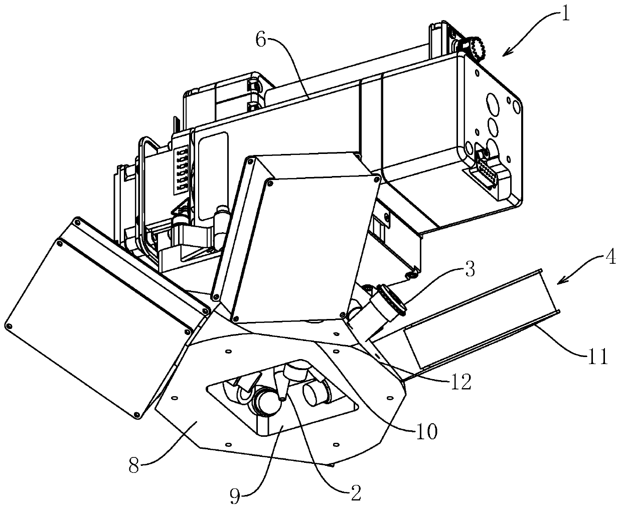
[0041] Such as figure 1 with figure 2 As shown, a multi-detector X-ray fluorescence micro-area scanning instrument includes an X-ray generating device 1, an X-ray converging device 2, an image acquisition device 3 for collecting image information, a detection device 4 for detecting fluorescence, and a sample moving device. Mobile device 5 and a computer for analyzing and processing data information.

[0042] Such as figure 2 As shown, the X-ray generating device 1 includes a housing 6 and a small focal spot X-ray tube located in the housing 6 . The X-ray converging device 2 is a polycapillary X-ray lens installed under the housing 6 and connected to the small focal spot X-ray tube. The X-rays generated by the small focal spot X-ray are converged by the polycapillary X-ray lens to form a minimum 5um The focal spot irradiates the sample, so that the intensity of the radiation received by the sample reaches more than 5000 times that when using a pinhole collimator.

[0043] S...

Embodiment 2

[0048] An imaging method of a multi-detector X-ray fluorescence micro-area scanning instrument, comprising:

[0049] S1. The X-ray generating device 1 generates X-rays to irradiate the sample, and the numerical control platform drives the sample to move at a set speed, and the numerical control platform sends the moving coordinate information to the computer at the same time.

[0050] S2. The four detectors 11 collect fluorescence signals, count them separately according to different energies / wavelengths, and send the detected data to the computer.

[0051] S3, the computer records the data measured by the four detectors 11 respectively, and adjusts the peak position data of the four detectors 11 based on any characteristic peak position, and sums up the adjusted data (automatically by computer, It can also be adjusted manually).

[0052] S4. Calculate the content of various elements in the sample at the injection focus position according to the summarized data, and combine t...


PUM


Abstract

The invention relates to the technical field of X-ray detection instruments, in particular to a multi-detector X-ray fluorescence micro-area scanning instrument and an imaging method thereof. An X-rayfluorescence micro-area scanning instrument with multiple detectors comprises an X-ray generating device, an X-ray converging device, a detecting device, a moving device and a computer for analyzingand processing data information, the detecting device comprises at least two detectors, and the distances between the detecting faces of all the detectors and the emitting focus of the X-ray converging device are equal. The X-ray generating device generates X-rays, and the generated X-rays are collected by the X-ray collecting device to form small focal spots. Fluorescence data generated after theX-ray irradiates the sample is collected by the detector and sent to the computer for analysis. The detection area and the counting rate are increased by arranging a plurality of detectors, and meanwhile, the dead time of the detectors is not increased, so that high-precision and high-efficiency scanning is realized.

Description

technical field [0001] The invention relates to the technical field of X-ray detection instruments, in particular to a multi-detector X-ray fluorescence micro-area scanning instrument and an imaging method thereof. Background technique [0002] XRF technology has the advantages of non-destructive, fast, wide analysis range, high quantitative accuracy, etc. It is widely used in metallurgy, building materials, mineral development and other fields. XRF quantitative range covers trace to constant, and there is no requirement for sample shape. It can complement other types of scanning techniques such as direct-reading spectroscopy and electron microscopy. In recent years, with the industrial upgrading of metallurgy, new material R&D and other fields and the continuous expansion of people's cognition to the microscopic field, the simple detection of the macroscopic element content of materials no longer meets the demand, and the concept of micro-scanning XRF has also come into bei...

Claims


Application Information

Patent Timeline
IPC IPC(8): G01N23/223G01N23/2204
CPCG01N23/2204G01N23/223G01N2223/3307
Inventor 刘明博赵雷胡学强杨博赞
Owner NCS TESTING TECH
Why Eureka
Try Eureka