Eureka AIR delivers breakthrough ideas for toughest innovation challenges, trusted by R&D personnel around the world.

i-motif recombination-mediated FRET probes and their application to in situ imaging of protein homodimerization on the surface of cancer cells

A homodimerization and protein technology, which is applied in material excitation analysis, fluorescence/phosphorescence, instruments, etc., can solve problems such as signal loss, and achieve high accuracy, universality, and high imaging fidelity

Active Publication Date: 2021-04-02
SHANDONG UNIV
View PDF12 Cites 0 Cited by
  • Summary
  • Abstract
  • Description
  • Claims
  • Application Information

AI Technical Summary

Problems solved by technology

This mismatch between FRET signal and protein homodimerization results in only half of the homodimeric proteins being detected by these methods, with severe loss of signal

Method used


Image

  • i-motif recombination-mediated FRET probes and their application to in situ imaging of protein homodimerization on the surface of cancer cells
  • i-motif recombination-mediated FRET probes and their application to in situ imaging of protein homodimerization on the surface of cancer cells
  • i-motif recombination-mediated FRET probes and their application to in situ imaging of protein homodimerization on the surface of cancer cells

Examples


preparation example Construction

[0042] The present invention also Provides a nucleic acid probe preparation method based on the i-motif recombination-mediated FRET strategy, including:

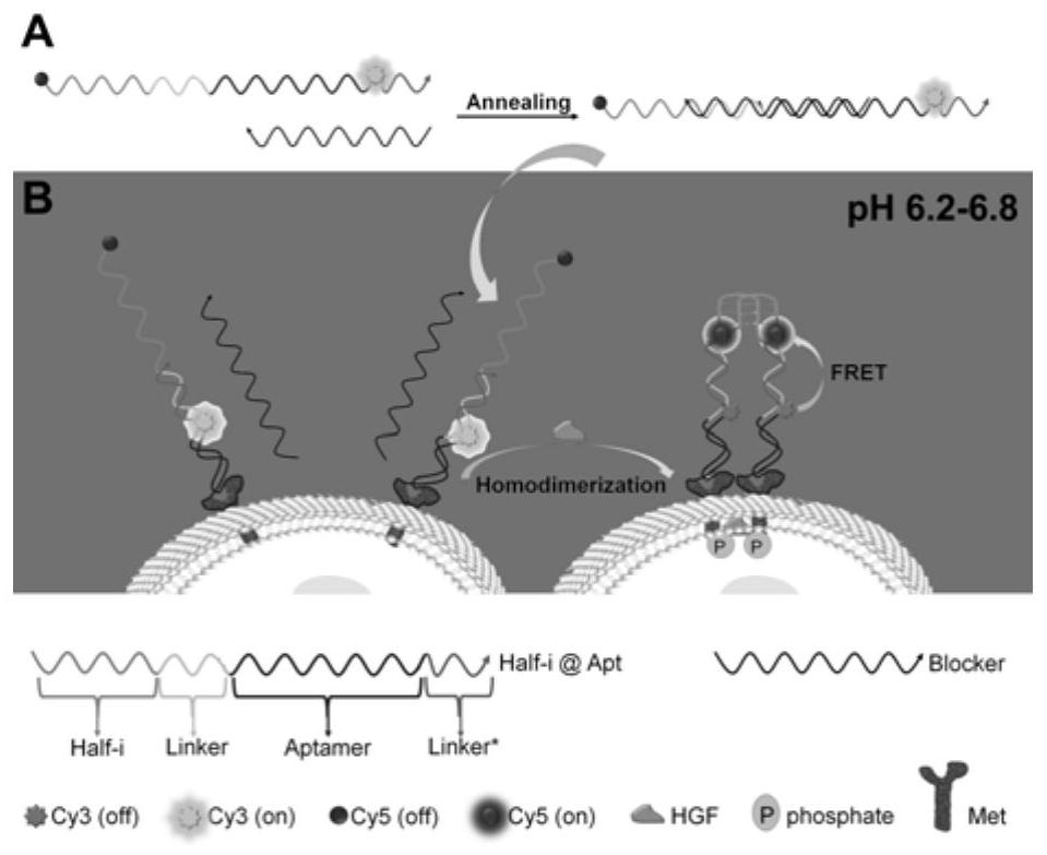
[0043] Mix the concentrated stock solutions of Half-i@Apt chains and Blocker chains, heat to 90-95°C for 4-5 minutes, then slowly cool to room temperature, and then place them in the dark at 3-5°C for more than 8 hours to make the two The strands are fully hybridized.

[0044] The probe preparation method of the present invention can adopt conventional preparation methods in the art to improve the preparation efficiency and the stability of the probe.

[0045] In some embodiments, the mixing ratio of the concentrated stock solutions of Half-i@Apt chains and Blocker chains is 1:1-1.5.

[0046] In some embodiments, the solvent of the concentrated stock solution is TE buffer to effectively dissolve the DNA powder.

[0047] The present invention also provides the application of any one of the above-mentioned probes in in s...

Embodiment 1

[0055] 1. Experimental part

[0056] 1.1 Materials and reagents

[0057] All DNA oligonucleotides were synthesized and purified by Sangon Bioengineering Co., Ltd. (Shanghai, China). DNA sequences and modifications are listed in Table 1. DMEM medium, RPMI 1640, fetal bovine serum (FBS), penicillin streptomycin (PS, 100U / ml), trypsin and phosphate-buffered saline (PBS, pH 7.4) for cell culture were all purchased from BiologicalIndustries (Israel ). Recombinant human hepatocyte growth factor (HGF, #100-39) was purchased from PeproTech (USA). Anti-HGF antibody [ab83760] was purchased from Abcam (UK). RIPA lysis buffer (P0013D), protease and phosphatase inhibitor cocktail (P1050) were purchased from Beyond Biotechnology Co., Ltd. (Shanghai, China). The primary antibody to β-Actin (AB0035) was purchased from Abways Technology (Shanghai, China). Primary antibodies to Met (#8198) and phospho-Met (Tyr1234 / 1235, #3077) were purchased from Cell Signaling Technology (USA). The seco...


PUM


Abstract

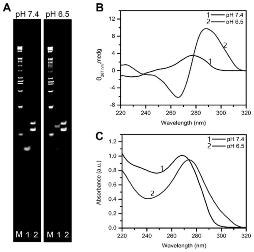
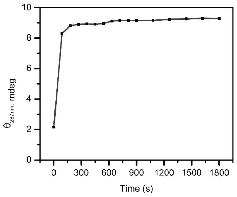
The invention relates to a i-motif recombination mediated FRET probe and an application of the i-motif recombination mediated FRET probe in in-situ imaging of cancer cell surface protein homodimerization. In the strategy, an aptamer of a target protein, half (Half-i) of an i-motif forming sequence and an FRET donor-receptor pair are integrated into the probe. The probe is bound to a target proteinthrough aptamer binding. The protein subjected to homodimerization leads the two probes to be close to each other, so the two same Half-i sequences can be recombined into i-motif in an acidic tumor extracellular environment, and the FRET effect is further caused. The matching degree of the FRET generation mode and the protein homodimerization mode in the design is higher. The strategy realizes accurate and dynamic in-situ imaging of mesenchymal epithelial transformation (Met) receptor homodimerization on the surfaces of tumor living cells, and has huge potential in biomedical research.

Description

technical field [0001] The invention belongs to the field of visual monitoring of protein homodimerization, and specifically relates to an i-motif recombination-mediated fluorescence resonance energy transfer (FRET) strategy for in situ imaging of homodimerization of surface proteins of living cancer cells . Background technique [0002] The information disclosed in this background section is only intended to increase the understanding of the general background of the present invention, and is not necessarily taken as an acknowledgment or any form of suggestion that the information constitutes the prior art already known to those skilled in the art. [0003] Membrane proteins perform most of their essential biological functions through protein-protein interactions. In particular, studies have identified the homodimerization of membrane proteins as an initial step in many signaling pathways that are critical in both physiological regulation and cancer progression. For examp...

Claims


Application Information

Patent Timeline
Patent Type & Authority Patents(China)
IPC IPC(8): G01N33/569G01N21/64
CPCG01N21/6428G01N33/56966G01N2333/71
Inventor 姜玮阚爱玲张楠
Owner SHANDONG UNIV
Why Eureka
Eureka Blog
Learn More