Fundus image segmentation method of full convolutional neural network based on Attention mechanism

A convolutional neural network and fundus image technology, applied in the field of medical image processing, can solve the problems of loss of useful information, inability to fully describe the optic disk of the fundus image, etc., and achieve the effect of improving accuracy and learning ability

Pending Publication Date: 2020-04-07
BEIJING INST OF OPHTHALMOLOGY +1
View PDF5 Cites 2 Cited by
  • Summary
  • Abstract
  • Description
  • Claims
  • Application Information

AI Technical Summary

Problems solved by technology

However, when using the existing fully convolutional neural network for fundus image segmentation, the process of extracting features layer by layer will cause a lot of useful information to be lost, which will lead to the fact that the parameters learned by the constructed model cannot fully describe the characteristics of the optic disc of the fundus image.

Method used


Image

  • Fundus image segmentation method of full convolutional neural network based on Attention mechanism
  • Fundus image segmentation method of full convolutional neural network based on Attention mechanism
  • Fundus image segmentation method of full convolutional neural network based on Attention mechanism

Examples


Embodiment Construction

[0046] The present invention is further described in conjunction with the following examples.

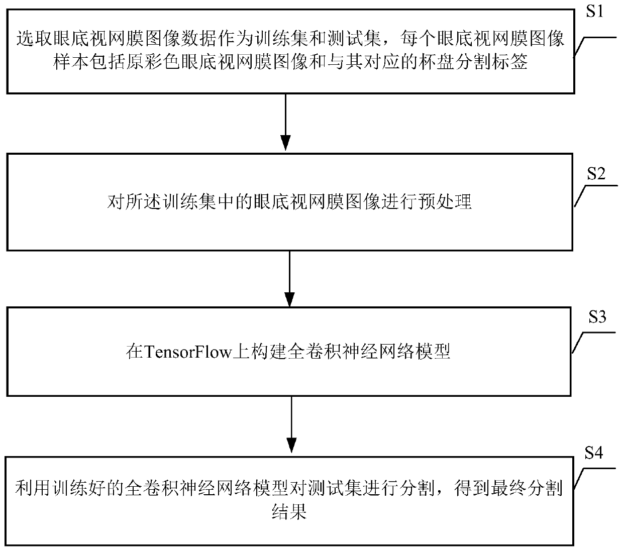
[0047] see figure 1 According to the first aspect, the embodiment of the present invention provides a fundus image segmentation method based on a fully convolutional neural network based on an Attention mechanism.

[0048] The method includes the following steps:

[0049] S1 selects the fundus retinal image data as the training set and the test set, and each fundus retinal image sample includes the original color fundus retinal image and its corresponding cup-and-disk segmentation label. Specifically, the cup-and-disc segmentation data of retinal images obtained from clinics are used as the training set to train the model, and the public 400 test data are used as the test set to test and evaluate the performance of the model.

[0050] S2 preprocesses the fundus retinal images in the training set, and uses them to input the fully convolutional neural network model. In an achievabl...


PUM


Abstract

The invention relates to the field of medical image processing, and provides a fundus image segmentation method and system of a full convolutional neural network based on an Attention mechanism, and acomputer readable storage medium, the method comprising: selecting fundus retina image data as a training set and a test set; preprocessing the fundus retina images in the training set; constructinga full convolutional neural network model on the TensorFlow; and segmenting the test set by using the trained full convolutional neural network model to obtain a final segmentation result. The systemcomprises a data acquisition module, a preprocessing module, a full convolutional neural network construction module and an image segmentation module. According to the multi-connection complete convolutional neural network model based on the Attention mechanism, the optic disc of the optic cup is automatically segmented from the fundus image, various limitations of a traditional method are overcome, the learning ability of the model is improved by fusing multi-level features in the neural network, and the accuracy of cup disc segmentation is improved.

Description

technical field [0001] The invention relates to the technical field of medical image processing, in particular to a fundus image segmentation method based on an Attention mechanism-based fully convolutional neural network. Background technique [0002] Optic cup and optic disc are one of the most basic organizational structures in retinal fundus images, and changes in the shape of the optic cup and optic disc are an important basis for clinical diagnosis of glaucoma. It is not only time-consuming and labor-intensive to manually segment fundus images to view the changes in the shape of the optic disc, but also the diagnostic results of different doctors are subjective to a certain extent, so it is not suitable for large-scale disease screening. Therefore, automatic retinal fundus image cup-disc segmentation is necessary to assist doctors in glaucoma screening. [0003] The existing methods for segmenting fundus images can be divided into traditional segmentation methods and ...

Claims


Application Information

Patent Timeline
Patent Type & Authority Applications(China)
IPC IPC(8): G06K9/00G06K9/34G06K9/62G06T7/00G06T7/10G06N3/04G06N3/08
CPCG06T7/0012G06T7/10G06N3/084G06T2207/30041G06T2207/10024G06T2207/20081G06T2207/20084G06V40/193G06V40/197G06V10/26G06V2201/03G06N3/045G06F18/253G06F18/214
Inventor 季鑫康宏
Owner BEIJING INST OF OPHTHALMOLOGY
Why Eureka
Try Eureka