Web-based chilled electron microscope data analysis graphical system and method
A cryo-electron microscope and data analysis technology, applied in image analysis, image data processing, image enhancement, etc., can solve time-consuming and energy-consuming problems, achieve the effects of reducing manpower, increasing segmentation speed, and reducing memory consumption
- Summary
- Abstract
- Description
- Claims
- Application Information
AI Technical Summary
Problems solved by technology
Method used
Image


Examples
Embodiment 1
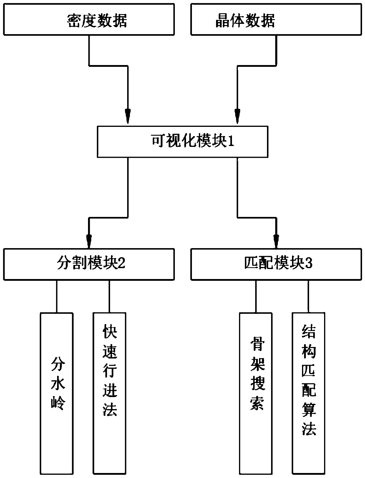
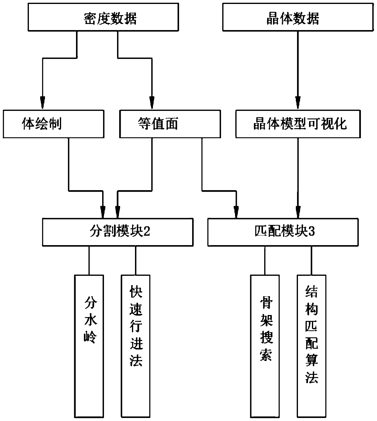
[0035] A web-based graphical system for cryo-EM data analysis, such as figure 1 As shown, including visualization module 1, segmentation module 2 and matching module 3, such as image 3 As shown, the visualization module 1 includes volume rendering, isosurface and crystal model visualization, and the cryo-electron microscopy data includes density data and crystal data; the visualization module 1 is used for visualizing the reconstructed density data and crystal structure data, and the volume The drawing and isosurface are used to display the density data, and the crystal data is displayed by the crystal model visualization module; the crystal data is used to match the density data in the later stage to derive the structure of the density data; the segmentation module 2 is used to analyze the three-dimensional data volume of the density data according to The characteristics of the macromolecular structure are segmented; the matching module 3 is used to match the density data wi...
Embodiment 2
[0053] As the second embodiment of the present invention, the multi-objective fast marching method includes initialization and circulation, and the specific steps of initialization are:
[0054] (1) mark the point whose radius is r on the axis of symmetry as the active point, let its time value T(x, y, z)=0;
[0055] (2) Mark the 6-connected neighbors of the active point as a close set, T(x, y, z)=1 / F(x, y, z);
[0056] (3) Mark all other points as far points, T(x,y,z)=TIME_MAX.
[0057] The specific steps of the cycle are:
[0058] (1) If the close collection is empty, exit the loop. Otherwise, select the point (i, j, k) with the smallest arrival time in the close set;
[0059] (2) Determine whether this point (i, j, k) is an active point of other partitions, if so, delete point (i, j, k) from the close set, and return to the above step (1);
[0060] (3) mark the point (i, j, k) as the active point, and delete it from the close collection;
[0061] (4) Mark the neighbors...
Embodiment 3
[0065] As the third embodiment of the present invention, HBV and RHDV are automatically segmented by the traditional fast-forward method and the fast-forward method of the present invention, and the memory test is carried out, as shown in the following table:
[0066] data number of blocks Legacy Data Structure (MB) This embodiment data structure (MB) HBV 42 1208 410 RHDV 32 796 345
[0067] It can be seen from the table that the new data structure can greatly reduce memory consumption.
[0068] The visualization module in this web-based cryo-electron microscope data analysis graphical system and method can display the data content, can visually see the data content, segment the entire three-dimensional data body according to the characteristics of the polyhedron structure, and realize the multi-objective fast marching method. The global segmentation of molecules improves the segmentation speed and reduces manpower for biologists to quickly ...
PUM
 Abstract
Description
Claims
Application Information
 - R&D
- Intellectual Property
- Life Sciences
- Materials
- Tech Scout
- Unparalleled Data Quality
- Higher Quality Content
- 60% Fewer Hallucinations
Browse by: Latest US Patents, China's latest patents, Technical Efficacy Thesaurus, Application Domain, Technology Topic, Popular Technical Reports.
© 2025 PatSnap. All rights reserved.Legal|Privacy policy|Modern Slavery Act Transparency Statement|Sitemap|About US| Contact US: help@patsnap.com