A diagnostic marker for bone destructive diseases and its application
A destructive and disease-related technology, applied in disease diagnosis, biological testing, biomaterial analysis, etc., can solve problems affecting the quality of life of patients
- Summary
- Abstract
- Description
- Claims
- Application Information
AI Technical Summary
Problems solved by technology
Method used
Image
Examples
Embodiment 1
[0056] Example 1 Changes in the expression of RNase L in the course of CIA rats
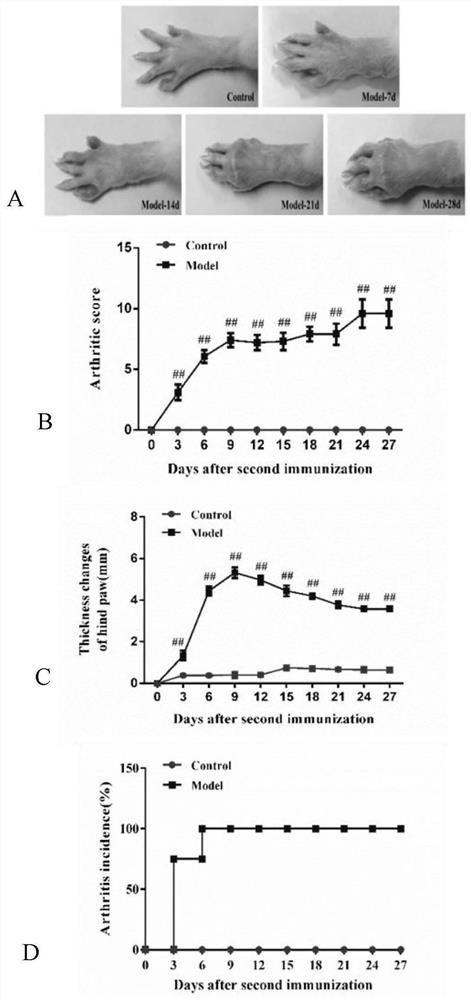
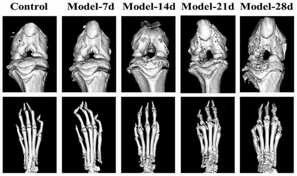
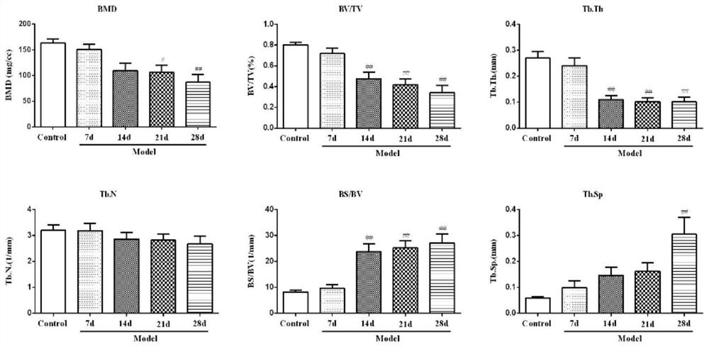
[0057] A total of 32 male SD rats with a body weight ranging from 160 to 180 g were selected, randomly divided into a blank control group (12 rats) and a model group (20 rats), and adaptively fed for 1 week. A collagen-induced arthritis (CIA) model was established one week later, and samples were collected on days 7, 14, 21, and 28 after the second immunization. Each sample included rats in the blank control group and the model group. All animals were fed a standardized diet and were not restricted in their activities.
[0058] 1. Effects on the clinical manifestations of CIA rats
[0059] On the 3rd day after the second immunization, the rats in the model group appeared symptoms of redness and swelling in the hindlimbs, which gradually involved the forelimbs. As time went by, the swelling of the knee joints of the hindlimbs gradually increased, and the degree of redness and swelling of the ankl...
Embodiment 2
[0064] Example 2 Effect of RNase L on the pathogenesis and bone destruction of CIA mice
[0065] Wild-type (WT) C57BL / 6 mice and RNase L gene knockout (RNase L - / - ) mice were randomly divided into 4 groups, namely WT-control (Control) group, WT-model (Model) group, RNase L - / - -Control group, RNase L - / - -Model group. In the Model group, the collagen-induced arthritis (CIA) model was established.
[0066] 1. Symptoms such as joint swelling and deformity
[0067] By establishing a CIA rat model, it was found that the expression of RNase L gradually increased over time. What is the effect of RNaseL on bone destruction in RA? The relationship between RNase L and RA was further clarified by establishing the CIA model of RNase L gene knockout mice. Redness and swelling of multiple interphalangeal joints appeared, and gradually spread to the foot pads and ankle joints, sometimes involving the forelimbs, and the degree of joint redness and swelling gradually decreased over time...
Embodiment 3
[0075] Example 3 Effect of RNase L on the pathogenesis and bone destruction of CAIA mice
[0076] CIA is an internationally recognized animal model of rheumatoid arthritis. In addition, we also adopted a collagen-antibody induced arthritis (CAIA) model, which is more clinical in its onset. C57BL / 6 WT mice and RNase L knockout mice were randomly divided into 4 groups, namely WT-Control group, WT-Model group, RNase L - / - -Control group, RNase L - / - -Model group. In the Model group, the type II collagen antibody-induced arthritis (CAIA) model was established.
[0077] 1. Effects on clinical manifestations of CAIA mice
[0078] On the 7th day after modeling, the mice in the model group began to have joint redness and swelling in the hind limbs, which gradually spread to the foot pads and ankle joints, and sometimes involved the forelimbs. In the later stage, the joint redness and swelling gradually reduced and symptoms of deformity and stiffness appeared. Under the same condit...
PUM
 Abstract
Description
Claims
Application Information
 - R&D
- Intellectual Property
- Life Sciences
- Materials
- Tech Scout
- Unparalleled Data Quality
- Higher Quality Content
- 60% Fewer Hallucinations
Browse by: Latest US Patents, China's latest patents, Technical Efficacy Thesaurus, Application Domain, Technology Topic, Popular Technical Reports.
© 2025 PatSnap. All rights reserved.Legal|Privacy policy|Modern Slavery Act Transparency Statement|Sitemap|About US| Contact US: help@patsnap.com


