Biological capsule for tissue repairing and establishing method thereof
A biocapsule and tissue repair technology, applied in the field of biomedical materials, can solve the problems of lack of inflammatory cell invasion and decomposition of biocapsules, and achieve good biocompatibility, prevent loss, enhance protection and mechanical strength
- Summary
- Abstract
- Description
- Claims
- Application Information
AI Technical Summary
Problems solved by technology
Method used
Image
Examples
Embodiment 1
[0033] Embodiment 1, the preparation of porous nanoscale honeycomb structure
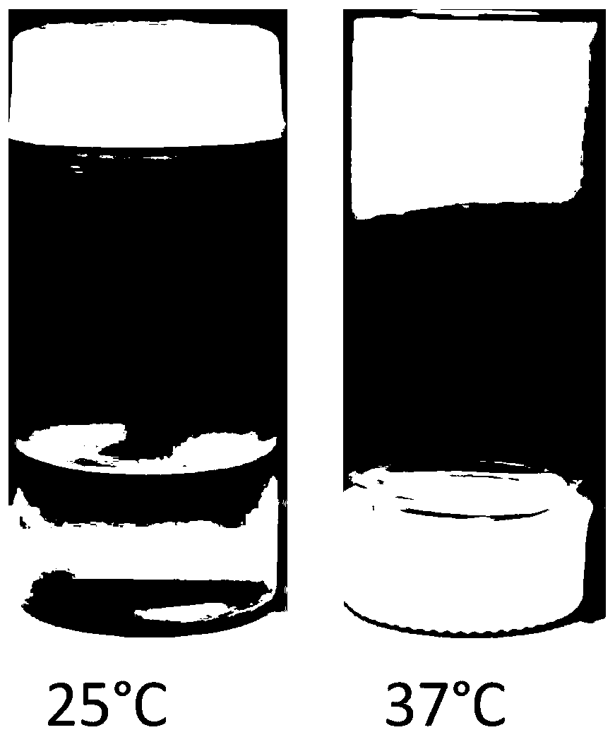
[0034] The porous nanoscale honeycomb structure is synthesized by free radical emulsion polymerization multipolymer, specifically, the preparation process is: 9.9mmol isopropylacrylamide NIPAM, 0.1mmol acrylic acid AA, 0.2mmol 2-mercaptobenzoic acid MBA and 0.2mmol Sodium dodecyl sulfate SDS was dissolved in 97 ml of water. After thorough mixing, the mixed solution was transferred to a 250ml three-necked flask equipped with a condenser and a mechanical stirrer.
[0035] The mixed solution was degassed under nitrogen for 30 min before polymerization. After degassing, the three-necked flask was placed in a preheated oil bath with a temperature of 70 °C. 3.0 ml of an aqueous KPS solution containing 0.1 mmol of potassium persulfate was injected into the mixture solution to initiate polymerization. Under the protection of nitrogen environment, the mixture solution was stirred continuously for 5 hours ...
Embodiment 2
[0036] Embodiment 2, porous nanoscale honeycomb structure
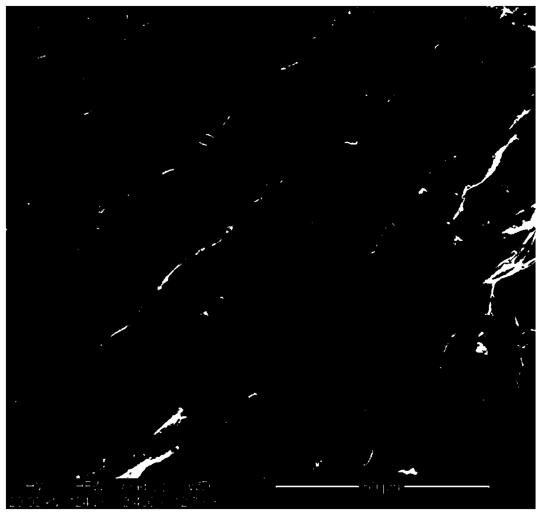
[0037] A scanning electron microscope (SEM) was used to observe the cross-section of the porous nano-scale honeycomb structure in the vertical and horizontal directions. Such as figure 2 As shown, the porous nano-scale honeycomb structure has a nano-scale pore size and can support or accommodate repair cells. The repair cells are cardiac stromal cells, mesenchymal stem cells or a combination thereof. Sources of repair cells include: tissue cells from the heart, mesenchymal stem cells from bone marrow and peripheral blood, etc. The porous nano-scale honeycomb structure is loaded with repair cells to form a biocapsule, which can better prevent the invasion of inflammatory cells, allow cells to grow better, and at the same time realize the release of small molecule bioactive substances.
Embodiment 3
[0038] Example 3, detection of internal morphology of biocapsules loaded with repaired cells
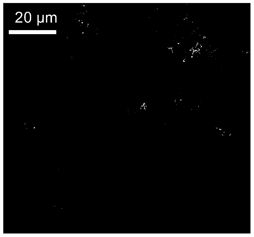
[0039] The repair cells and cardiac stromal cells were collected in the culture medium and mixed with 10XPBS and the liquid of the porous nanoscale honeycomb structure at a ratio of 1:1:3, and then heated in a 37°C incubator to gel to form biological capsule.
[0040] Observe the biocapsules with a scanning electron microscope. Specifically, the biocapsules are dehydrated and fixed, encapsulated on an aluminum post with double-sided tape and coated with a thin layer of gold. Under an accelerating voltage of 15kV, the biocapsules are scanned and photographed by a scanning electron microscope. Such as image 3 As shown, the results of the biocapsules indicated that the porous nanoscale honeycomb structure encapsulated the repair cells.
PUM
 Abstract
Description
Claims
Application Information
 - R&D
- Intellectual Property
- Life Sciences
- Materials
- Tech Scout
- Unparalleled Data Quality
- Higher Quality Content
- 60% Fewer Hallucinations
Browse by: Latest US Patents, China's latest patents, Technical Efficacy Thesaurus, Application Domain, Technology Topic, Popular Technical Reports.
© 2025 PatSnap. All rights reserved.Legal|Privacy policy|Modern Slavery Act Transparency Statement|Sitemap|About US| Contact US: help@patsnap.com


