


Quick Research
Generate reliable direction feasibility study reports for your R&D in just a few steps.

Technical Q&A
Discover and master advanced knowledge NOW. Basics, ideas, possibilities, all at once.

Find Solutions
As an expert in R&D theories, this can generate solutions to your technical problems instantly.

Evaluate Feasibility
Analyze your overall solution with one click, know your potential R&D risks in advance.

Monitor Landscape
Get weekly tech updates, stay abreast of the latest tech innovations and key insights.
Method for measuring human cartilage endochrome and application of human cartilage endochrome in forensic medicine
A method of measuring cartilage, which is applied in the field of forensic anthropology, can solve the problems of inability to obtain the objective relationship of cartilage pigmentation, inaccurate quantification, and cumbersome operations
- Summary
- Abstract
- Description
- Claims
- Application Information
AI Technical Summary
Problems solved by technology
Method used
Image

Examples
Embodiment 1
[0048] Example 1 Measuring method of pigment in costal cartilage
[0049] (1) Cost cartilage sample processing and image acquisition
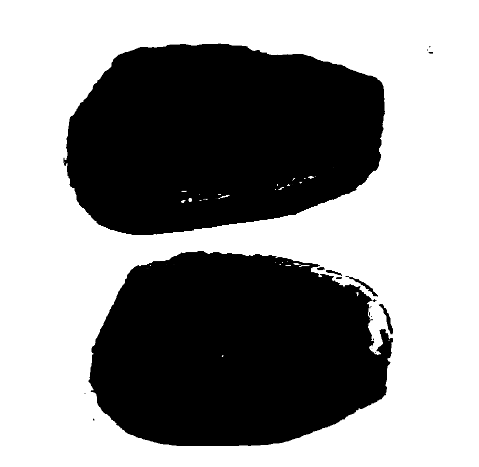
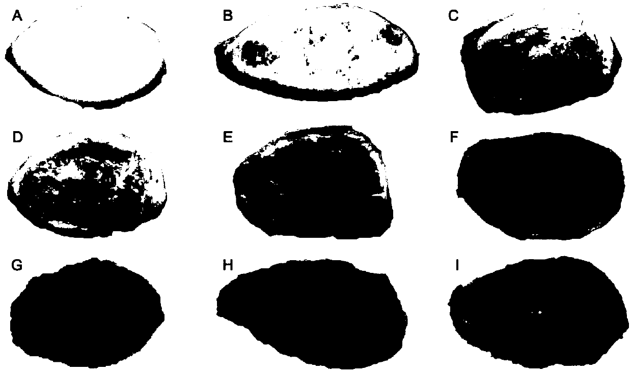
[0050] In this embodiment, the anterior ribs of the second costal cartilage isolated from 89 Han people (including 58 men and 31 women) were used to obtain tissue sections of the second costal cartilage. Remove the extraneous attached tissue of the isolated cartilage to avoid damaging the cartilage tissue itself, wash it with water and dry it. Use a scalpel or other slicing tool to obtain a slice of cartilage tissue with a thickness of about 2 mm through a cross-sectional incision. Use Canon (Canon) 9000F Mark Ⅱ scanner "photo scan mode" to scan the two sides of costal cartilage slices to obtain a color image with an image format of TIFF and an image resolution of DPI=600.
[0051] (2) Obtaining the average gray value of the costal cartilage section image
[0052] The image analysis software Adobe Photoshop CS5 was used to read the image of the costa...
Embodiment 2
[0053] Example 2. Analysis of correlation between costal cartilage pigment and age
[0054] (1) The establishment of a mathematical model for age estimation based on quantification of pigment in costal cartilage
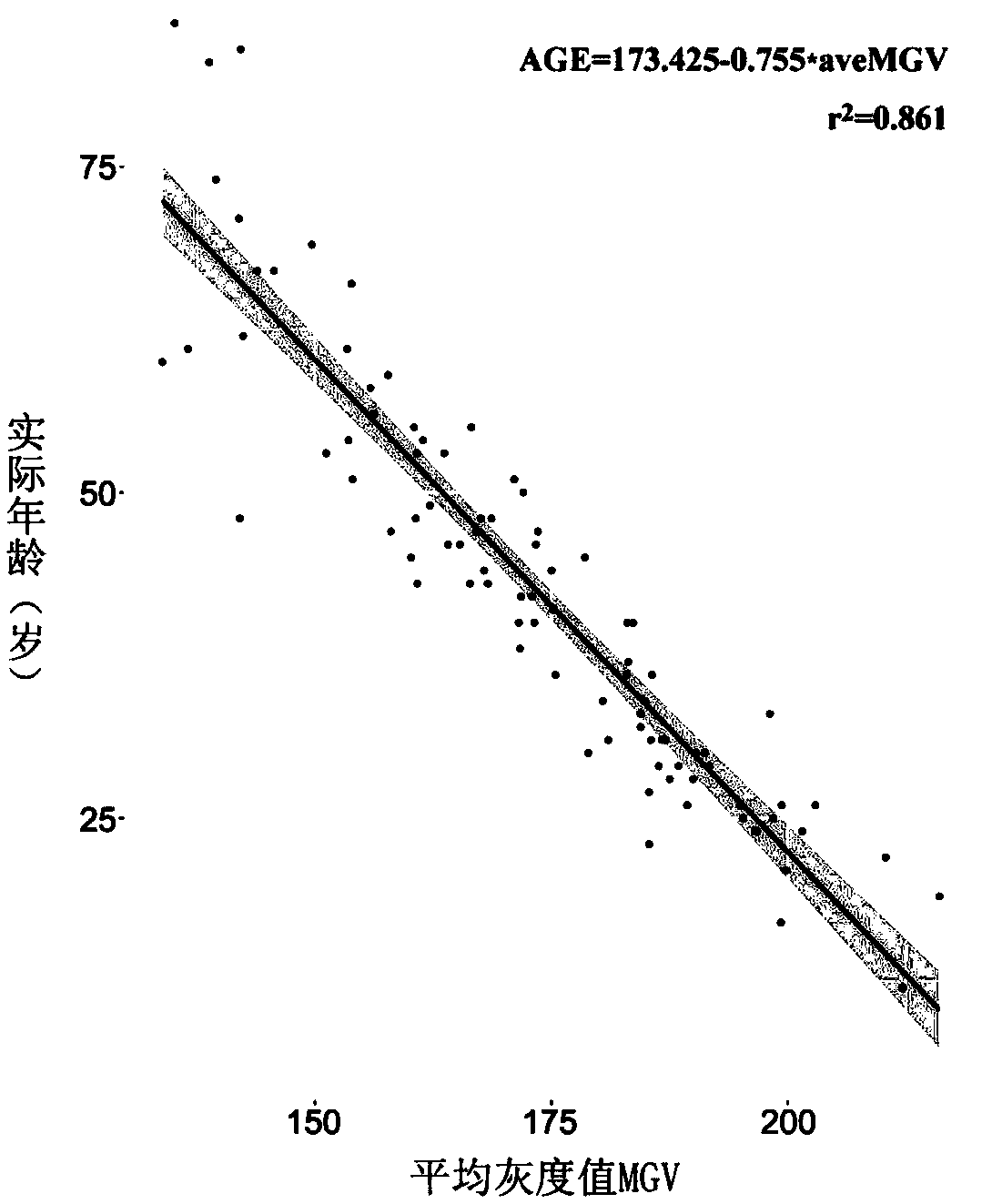
[0055] The age and gender of the 89 samples were used as independent variables, and the average gray value of the corresponding costal cartilage section was used as the dependent variable. The variables were analyzed by multiple linear regression model using SPSS software. In the Pearson correlation analysis, the age variable was significantly correlated with the average gray value of the costal cartilage section (p 0.05), indicating that there is no significant difference in the amount of pigment in the costal cartilage between men and women.
[0056] The SPSS software was used to analyze the age and the average gray value of costal cartilage section with a univariate linear regression model. Among them, age is used as the dependent variable, and the average gray value o...
Embodiment 3
[0059] Example 3 Application of quantification of intrachondral pigment in forensic medicine (verification of the accuracy of the age estimation model based on quantification of human costal cartilage pigment)
[0060] Collect the second cost cartilage in vitro from 24 Chinese Han individuals, and obtain the second cost cartilage slices from the body at a distance of 2 cm from the sternum according to the measurement method in Example 1 above, and obtain the scan pictures of each cost cartilage slice. And the average gray value. The average gray value of each costal cartilage section is substituted into the age estimation model obtained in Example 2, and the estimated age is calculated. The estimated age is compared with the actual age, and the average absolute age deviation (MAD) is calculated. The specific results are shown in the following table:
[0061]
[0062]
[0063] The results showed that the accuracy of the age estimation model based on quantification of the pigment in...
PUM
| Property | Measurement | Unit |
|---|---|---|
| Slice thickness | aaaaa | aaaaa |
Abstract
Description
Claims
Application Information
 - R&D Engineer
- R&D Manager
- IP Professional
- Industry Leading Data Capabilities
- Powerful AI technology
- Patent DNA Extraction
Browse by: Latest US Patents, China's latest patents, Technical Efficacy Thesaurus, Application Domain, Technology Topic, Popular Technical Reports.
© 2024 PatSnap. All rights reserved.Legal|Privacy policy|Modern Slavery Act Transparency Statement|Sitemap|About US| Contact US: help@patsnap.com