Capsule endoscope

A technology of capsule endoscope and capsule shell, applied in the fields of endoscopy, medical science, diagnosis, etc., can solve the problem that capsule power supply efficiency and image transmission efficiency cannot be fully guaranteed, reduce the working performance of capsule endoscope system, capsule The problem of low utilization rate of endoscope power supply can achieve the effect of improving clinical detection rate, improving efficiency and increasing utilization rate

Inactive Publication Date: 2019-08-23
WUXI ANZHIZHUO MEDICAL ROBOT CO LTD
View PDF0 Cites 1 Cited by
  • Summary
  • Abstract
  • Description
  • Claims
  • Application Information

AI Technical Summary

Problems solved by technology

[0005] At present, the power supply of the capsule endoscope will always be in the working state after it is turned on, which causes the power utilization rate of the capsule endoscope to be low. The capsule endoscope, which is mainly used to inspect the small intestine or large intestine, will be consumed in the esophagus and stomach. A large amount of power supply power, which will cause clinical missed detection of small intestine and large intestine examination, and reduce the performance of the product
At the same time, the capsule endoscope moves and works in the organs of the digestive tract. Due to the different structures of different organs and tissues and the harsh internal environment, the power supply efficiency and image transmission efficiency of the capsule cannot be fully guaranteed, which ultimately reduces the performance of the capsule endoscope as a whole. The performance of the mirror system

Method used


Image

  • Capsule endoscope

Examples


Embodiment Construction

[0020] In order to make the objects and advantages of the present invention clearer, the present invention will be further described in detail below in conjunction with the examples. It should be understood that the specific embodiments described here are only used to explain the present invention, not to limit the present invention.

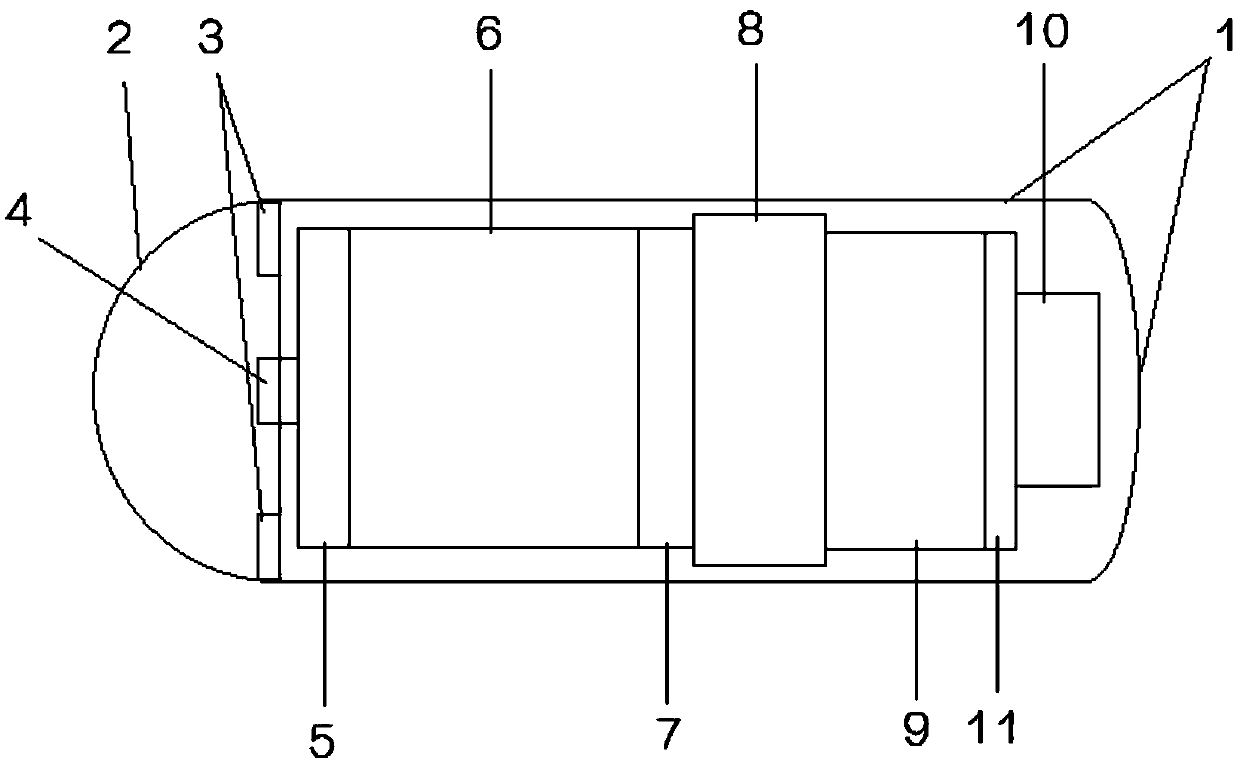
[0021] Such as figure 1 As shown, the embodiment of the present invention provides a capsule endoscope, including a capsule shell 1 and an image acquisition device disposed inside the capsule shell 1, a main control circuit module 6, a power management module 11, a battery 8, and a wireless transceiver module 9 1. Antenna 10; the capsule shell 1 is smooth cylindrical, and one end thereof is provided with an optical window 2; the optical window 2 is smooth and transparent hemispherical, and the battery 8 is a silver oxide button battery.

[0022] The image acquisition device is used to collect image data in the subject, and transmits the image d...


PUM


Abstract

The invention discloses a capsule endoscope which comprises a capsule shell, an image acquisition device, a main control circuit module, a power management module, a battery, a wireless transceiving module and an antenna, wherein the image acquisition device, the main control circuit module, the power management module, the battery, the wireless transceiving module and the antenna are arranged inside the capsule shell; the image acquisition device is used for acquiring image data in a detected body and transmitting the image data to the wireless transceiving module through the main control circuit module; the wireless transceiving module comprises a wireless chip and a peripheral circuit; the antenna is connected with the wireless transceiving module and comprises a flexible antenna and abracket used for transmitting the image data received by the wireless transceiving module. A power supply is regularly controlled through the power supply management module, the utilization rate of the battery is improved to the maximum extent, the power supply management module of the capsule endoscope can be controlled according to an instruction of an external receiver, the performance of the battery is fully and effectively utilized, the working time of the capsule endoscope is prolonged, the digestive tract can be fully covered and examined, and the overall working performance of the capsule endoscope is improved.

Description

technical field [0001] The invention relates to the technical field of medical instruments, in particular to a capsule endoscope. Background technique [0002] At present, after the subject swallows the capsule endoscope, due to the natural peristalsis of the digestive tract organs, the capsule can automatically move and send the collected images to the outside of the body, and the receiver outside the body can display the image in real time after receiving the image data , At the same time, the pictures can also be saved in the memory. After the examination, the doctor can read the image data through the workstation and make a medical diagnosis based on the image. [0003] Israel's Given Imaging company took the lead in launching the M2A capsule endoscope, and soon obtained the US FDA certification and entered clinical use. Subsequently, Given Imaging successively launched the Pillcam series of capsule endoscopes for the inspection and diagnosis of the esophagus, small int...

Claims


Application Information

Patent Timeline
IPC IPC(8): A61B1/04A61B1/06
CPCA61B1/00032A61B1/00043A61B1/041A61B1/0684
Inventor 王海涛
Owner WUXI ANZHIZHUO MEDICAL ROBOT CO LTD
Why Eureka
Try Eureka