Fluorescent OCT dual-mode imaging method and device based on indocyanine green nanometer

An indocyanine green nanometer and dual-mode imaging technology, which is applied in the fields of fluorescence/phosphorescence, material excitation analysis, etc., can solve the problem of not being able to explore the signal in the depth direction, and achieve the effect of rapid detection and response

Active Publication Date: 2019-05-24
FOSHAN UNIVERSITY
View PDF14 Cites 3 Cited by
  • Summary
  • Abstract
  • Description
  • Claims
  • Application Information

AI Technical Summary

Problems solved by technology

However, at present, ICG imaging is generally realized by using fluorescence imaging technology, and fluorescence imaging only obtains the surface information of the detection area, and cannot explore the shortcomings of the depth direction signal.

Method used


Image

  • Fluorescent OCT dual-mode imaging method and device based on indocyanine green nanometer
  • Fluorescent OCT dual-mode imaging method and device based on indocyanine green nanometer
  • Fluorescent OCT dual-mode imaging method and device based on indocyanine green nanometer

Examples


Embodiment Construction

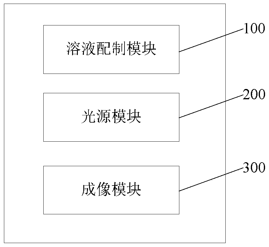
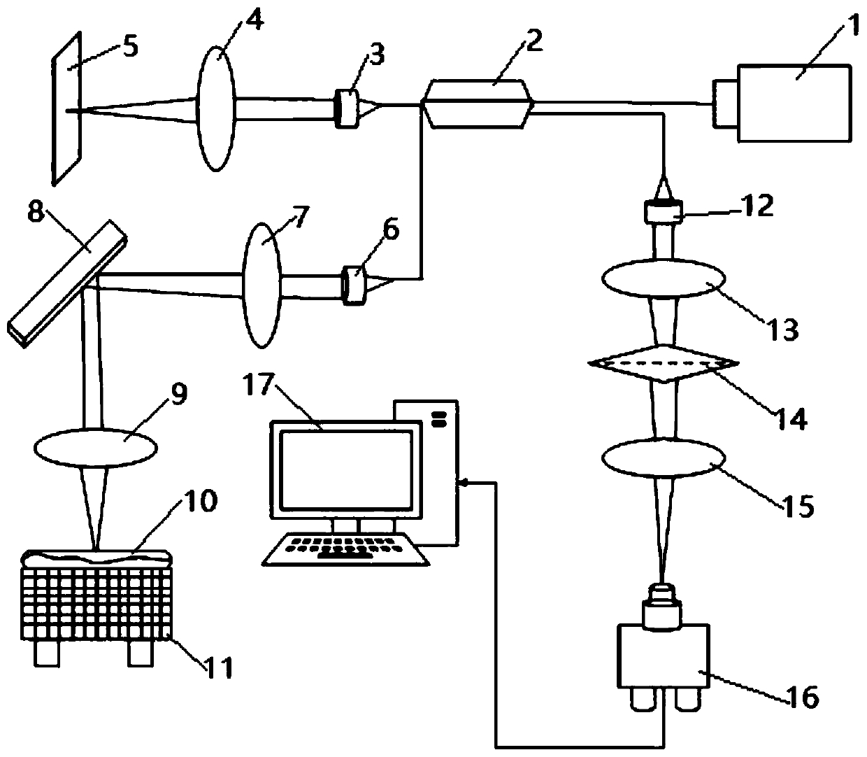
[0041] Below in conjunction with accompanying drawing, the present invention will be further described. In order to obtain more biological information at the same time, the present invention uses indocyanine green nanomaterials as a contrast agent to combine the two imaging methods of fluorescence imaging and OCT imaging into a new imaging system—fluorescence-OCT dual-mode imaging system. The dual-mode imaging system can image the position and depth of the specific biological lesion area, provide accurate sample information, and improve the specific diagnosis of certain diseases, which has potential application value in medicine. In some implementations, according to the light absorption or scattering characteristics of different imaging targets, light sources with different wavelengths can be selected to simultaneously obtain the functional and structural imaging information of the object within a certain depth range, and finally the specific location of the lesion can be clea...


PUM


Abstract

The invention discloses a fluorescent-OCT dual-mode imaging method and device based on indocyanine green nanometer. The method includes: compounding indocyanine green with polyethylene glycol after being treated with phospholipid to form indocyanine green-phospholipid-treated polyethylene glycol nanoparticles, and modifying biologically targeted molecules at the tail ends of polyethylene glycol molecules to obtain an ICG-PL-PEG-FA solution; injecting the ICG-PL-PEG-FA solution to an imaging target, utilizing a low-coherence light source of near infrared to irradiate a to-be-scanned area of theimaging target, and using a light source of 780nm in wavelength to excite ICG-PL-PEG-FA molecules in the imaging target to generate fluorescence; opening an OCT imaging light path to acquire OCT of the imaging target to develop a structure image with a depth range. The fluorescence is generated through the ICG-PL-PEG-FA molecules in the imaging target, an OCT imaging system is applied to collectfluorescent signals, and position information of fluorescent substances in an experimental living body can be reduced, so that lesion area position recognized by a contrast agent can be clearly distinguished, and the structure image of a certain depth range can be presented.

Description

technical field [0001] The invention belongs to the field of biological imaging, in particular to a fluorescence-OCT dual-mode imaging method and device based on indocyanine green nanometers. Background technique [0002] Optical coherence tomography (OCT) and fluorescence imaging technology are widely used in medical diagnosis. OCT technology uses the basic principle of weak coherent light interference to detect the back reflection or several times of incident weak coherent light at different depths of biological tissue. Scattering signals, through scanning, can obtain biological tissue depth signals, reconstruct two-dimensional or three-dimensional structural images, and thus obtain biological depth information. Fluorescence imaging technology uses fluorescent probes to generate and collect fluorescent signals by exciting the probes with an excitation light source. A single program mode cannot obtain comprehensive functional and structural imaging information to understan...

Claims


Application Information

Patent Timeline
IPC IPC(8): G01N21/64
Inventor 钟俊平黎思娜曾亚光韩定安岑臻涛罗梦诗
Owner FOSHAN UNIVERSITY
Why Eureka
Try Eureka