Method for quantitatively evaluating myocardial infarction on basis of nuclide image and CT (computed tomography) coronary angiography fusion

A technique for quantitative assessment of myocardial infarction, applied in the field of medical image processing, can solve problems such as complex heart image registration, different heart image scanning time, and different scanning equipment, so as to avoid interference from human factors, high degree of automation, and matching The effect of high quasi-efficiency

Inactive Publication Date: 2019-03-22
XIDIAN UNIV
View PDF5 Cites 10 Cited by
  • Summary
  • Abstract
  • Description
  • Claims
  • Application Information

AI Technical Summary

Problems solved by technology

However, the scanning time of cardiac images of different modalities is different, the scanning equipment is different, the patient's heart rate, blood flow and other factors will cause the difference between the images of different modalities, and the non-rigid deformation of the heart makes different modalities The problem of cardiac image registration in the state becomes very complex

Method used


Image

  • Method for quantitatively evaluating myocardial infarction on basis of nuclide image and CT (computed tomography) coronary angiography fusion
  • Method for quantitatively evaluating myocardial infarction on basis of nuclide image and CT (computed tomography) coronary angiography fusion
  • Method for quantitatively evaluating myocardial infarction on basis of nuclide image and CT (computed tomography) coronary angiography fusion

Examples


Embodiment Construction

[0042] In order to make the object, technical solution and advantages of the present invention more clear, the present invention will be further described in detail below in conjunction with the examples. It should be understood that the specific embodiments described here are only used to explain the present invention, not to limit the present invention.

[0043] The application principle of the present invention will be described in detail below in conjunction with the accompanying drawings.

[0044] Such as figure 1 As shown, the concrete steps of the myocardial infarction quantitative assessment method based on nuclide image and CT coronary angiography fusion of the present invention are as follows:

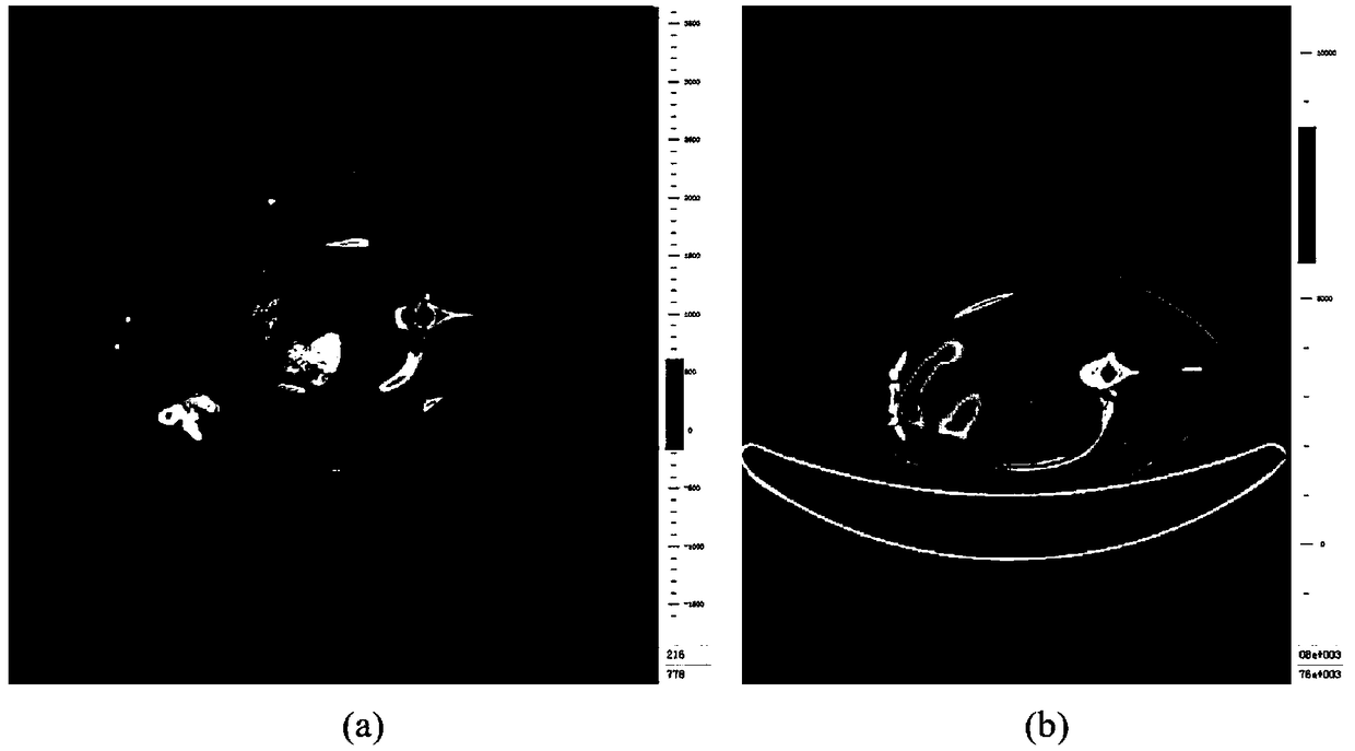
[0045] Step 1. Input cardiac CT coronary angiography image and cardiac nuclide image, adjust image pixel size:

[0046] Cardiac CT coronary angiography images and heart images are obtained from a hospital, such as figure 2 shown, where figure 2 (a) is a cardiac CT corona...


PUM


Abstract

The invention belongs to the field of medical image processing, and discloses a method and a system for quantitatively evaluating myocardial infarction on the basis of nuclide image and CT (computed tomography) coronary angiography fusion. The method includes acquiring heart PET (positron emission tomography) / CT and CTA (computed tomography angiography) data; carrying out three-dimensional DCT (discrete cosine transformation) frequency-domain interpolation on PET / CT images; acquiring heart regions in the PET / CT images and CTA images by the aid of multi-spectrum segmentation processes; acquiring left ventricular cardiac muscle regions in PET images by the aid of threshold processes; manually segmenting left ventricular cardiac muscle regions in the CTA images layer by layer; registering images; classifying pixel points according to the locations of pixel points on the registered images and tracer uptake values; inversely transforming infarction regions into original CTA spaces and dividing the volumes of left ventricular cardiac muscles from the volumes of the transformed infarction regions; three-dimensionally displaying fused images in volume rendering manners. The method and thesystem have the advantages that integral registration procedures can be fully automated, the method and the system are good in objectivity, intuitive results can be obtained, and analysis can be facilitated for clinical doctors.

Description

technical field [0001] The invention belongs to the field of medical image processing, and in particular relates to a quantitative evaluation method and evaluation system for myocardial infarction based on the fusion of nuclide images and CT coronary angiography. Background technique [0002] At present, the existing technology commonly used in the industry is as follows: Myocardial infarction is a kind of disease that causes myocardial necrosis due to coronary ischemia and hypoxia, and eventually leads to sudden death (acute) and myocardial remodeling (chronic), which seriously threatens human life and affects Quality of Life. Infarct size is a decisive factor for myocardial remodeling and cardiac dysfunction, and is an effective predictor of survival in patients with myocardial infarction. After myocardial infarction, the accurate positioning and quantification of the ischemic area will help clinically to effectively open the infarction-related coronary arteries and save ...

Claims


Application Information

Patent Timeline
Patent Type & Authority Applications(China)
IPC IPC(8): A61B6/03
CPCA61B6/032A61B6/037A61B6/4417A61B6/503A61B6/5217A61B6/5235
Inventor 梁继民徐真真陶博曹丰任胜寒陈雪利胡海虹
Owner XIDIAN UNIV
Why Eureka
Try Eureka