
A brain MRI image segmentation method combined with biological characteristics

An image segmentation and biometric technology, applied in the field of image processing and biomedicine, can solve problems such as a large number of training sets, and achieve the effect of solving training samples, improving accuracy, and improving accuracy.

Inactive Publication Date: 2019-03-08
YANCHENG CHITTAGONG SMART TERMINAL IND RES INST CO LTD
View PDF3 Cites 2 Cited by
  • Summary
  • Abstract
  • Description
  • Claims
  • Application Information

AI Technical Summary

Problems solved by technology

[0004] In view of the fact that the current segmentation methods do not combine brain structural features, and the machine learning method requires a large number of training sets, the present invention provides an unsupervised random forest brain MRI image segmentation method combined with the biological feature structure of cerebrospinal fluid

Method used


Image

  • A brain MRI image segmentation method combined with biological characteristics
  • A brain MRI image segmentation method combined with biological characteristics
  • A brain MRI image segmentation method combined with biological characteristics

Examples


Embodiment Construction

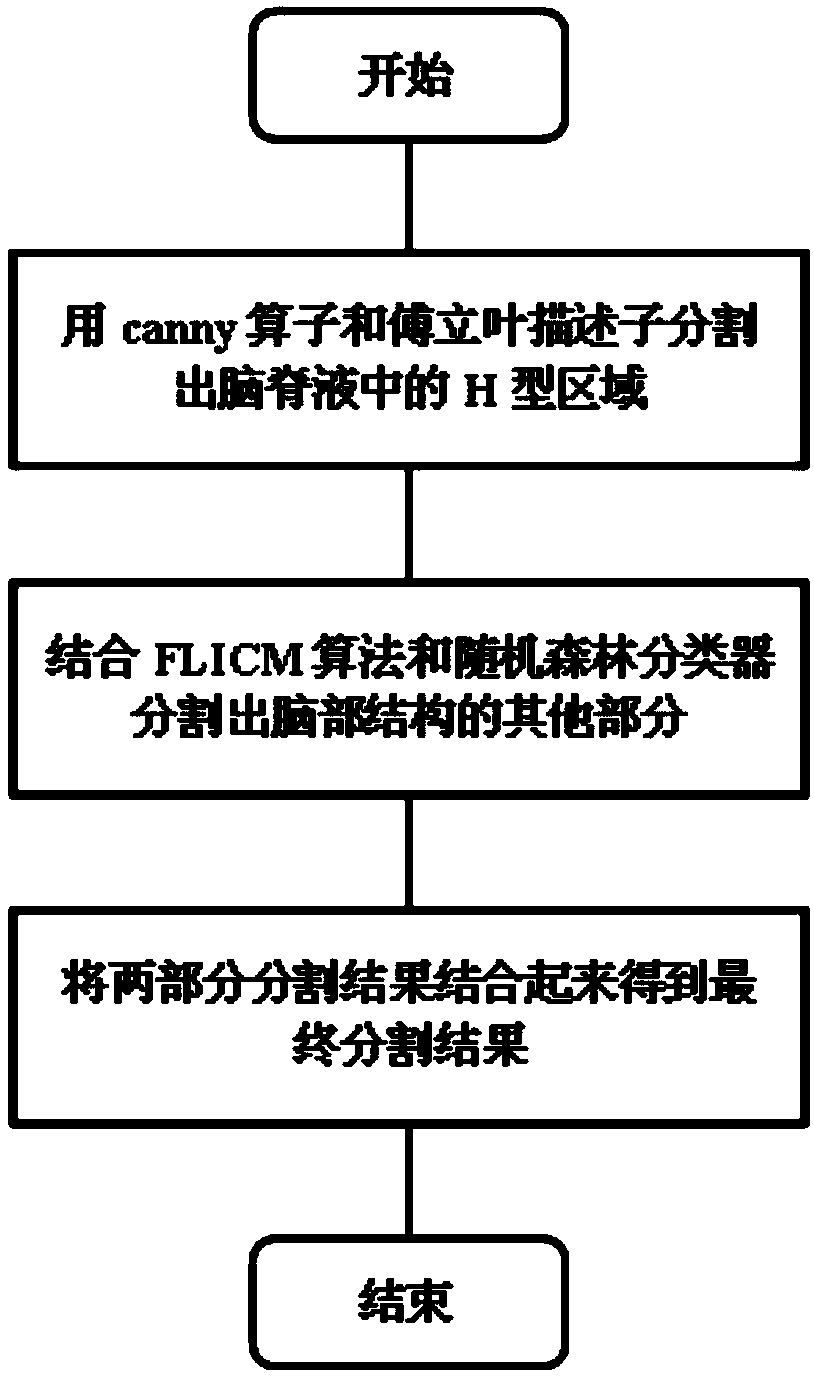
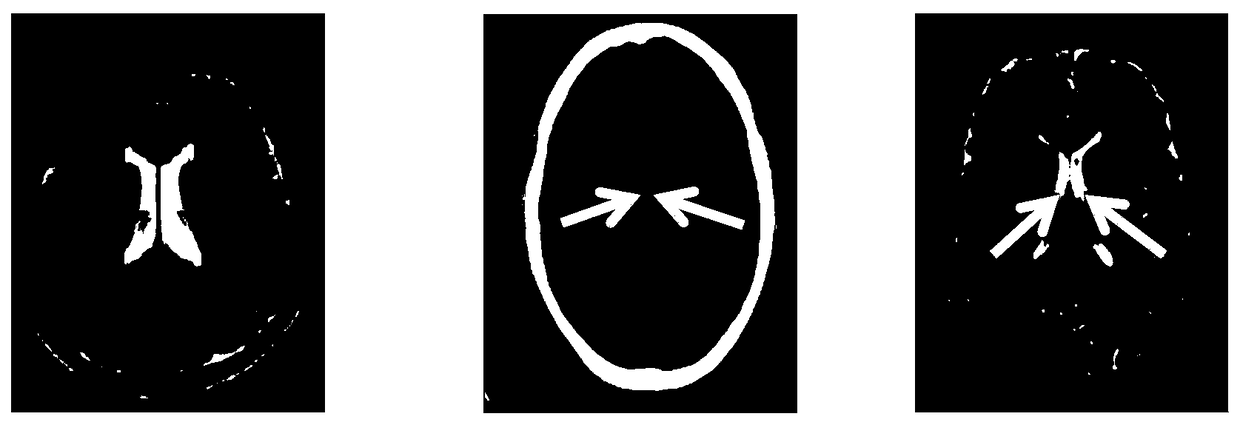
[0044] The core content of the present invention is that: in the segmentation process of the MRI image, the biological feature that the central part of the cerebrospinal fluid is H-shaped is utilized, thereby improving the segmentation accuracy of the cerebrospinal fluid. And the combination of FLICM algorithm and random forest classifier realizes complete unsupervised segmentation.

[0045] In order to make the object of the present invention, technical scheme and advantage clearer, do further detailed description below in conjunction with accompanying drawing and example:

[0046] 1. Segmenting the central H-shaped region of the cerebrospinal fluid in the MRI image includes the following steps:

[0047] 1.1 Obtain brain MRI images from the brainweb database;

[0048] 1.2 First use the canny operator to extract the edge of the H-shaped area in the standard MRI cerebrospinal fluid segmentation map, and then perform discrete Fourier transform on it. The coefficient of the disc...


PUM


Abstract

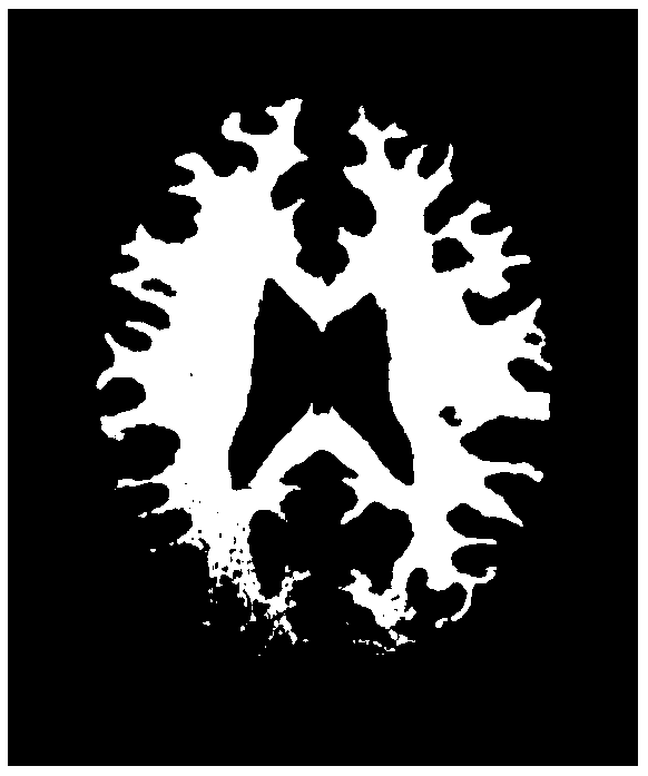
The invention relates to a brain MRI image segmentation method combined with biological characteristics, belonging to the technical field of image processing and biomedical combination. The method ofthe present invention utilizes H-type biometric features of CSF central part to segment CSF part, the rest of the brain is segmented by the fuzzy C-means clustering algorithm based on local information as the initial label input of the random forest, the symmetry of the brain image is used, the left half pixels of the MRI image are used as the training samples of the random forest classifier, andall the pixels are used as the test samples of the random forest classifier. Finally, the final segmentation result of the brain MRI image is obtained by combining the segmentation results of the twoparts. The final result of the invention is better than other segmentation algorithms only considering image gray information, and the unsupervised segmentation without requiring a large number of picture training sets is realized.

Description

technical field [0001] The invention belongs to the technical field of combining image processing and biomedicine, and in particular relates to a brain MRI image segmentation method combined with biological features. Background technique [0002] MRI is a type of tomographic imaging, which uses the phenomenon of magnetic resonance to obtain electromagnetic signals from the human body and reconstruct the information of the human body. Since MRI can image without injecting radioactive isotopes, it is safer and has higher resolution than imaging techniques such as CT and PET. Nowadays, computer-aided medical diagnosis technology has been very popular. For the analysis and diagnosis of computer-aided brain MRI images, the most critical step is image segmentation. The accuracy of segmentation will directly affect the follow-up diagnosis work. The brain structure of the human body is mainly composed of three parts: white matter, gray matter, and cerebrospinal fluid. [0003] At ...

Claims


Application Information

Patent Timeline
Patent Type & Authority Applications(China)
IPC IPC(8): G06T7/12G06T7/13G06T7/168G06T7/136G06T7/11G06K9/62
CPCG06T7/11G06T7/12G06T7/13G06T7/136G06T7/168G06T2207/20056G06T2207/20081G06T2207/30016G06T2207/10088G06F18/23213G06F18/24323
Inventor 赵岩李梓萌王世刚
Owner YANCHENG CHITTAGONG SMART TERMINAL IND RES INST CO LTD
Learn More