Cell capture and fluorescence enhancement device and method based on biological lens
A fluorescence enhancement and lens technology, applied in the field of optics, can solve the problems that up-conversion fluorescent nanoparticles cannot achieve high biocompatibility, high efficiency, high precision and high flexibility, expensive devices, complex structures, etc. Small limitations, improved luminous efficiency, and small volume effects
- Summary
- Abstract
- Description
- Claims
- Application Information
AI Technical Summary
Problems solved by technology
Method used
Image
Examples
Embodiment 1
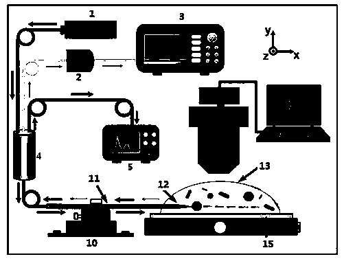
[0069] The method for cell capture and fluorescence enhancement based on a biolens is specifically performed according to the following steps:
[0070] Step S1: Prepare an optical fiber probe 12 for capturing experimental objects and detecting signals. The optical fiber probe 12 is prepared from a standard multimode optical fiber through the fusion tapered method, specifically according to the following steps:
[0071] Step S11, after stripping off a 2 cm coating layer in the middle of the multimode optical fiber with optical fiber wire strippers, insert it into the capillary glass tube 11 to protect the multimode optical fiber. The inner diameter of the capillary glass tube 11 is 0.9 mm, and the wall 0.1 mm thick and 12 cm long;
[0072] Step S12, place the bare multimode optical fiber in parallel on the outer flame above the alcohol lamp, let it stand for 1.5 minutes until the optical fiber is melted, and draw the melted part with the help of hands at a speed of 6 mm per sec...
Embodiment 2
[0083] The method for cell capture and fluorescence enhancement based on a biolens is specifically performed according to the following steps:
[0084] Step S1: Prepare an optical fiber probe 12 for capturing experimental objects and detecting signals. The optical fiber probe 12 is prepared from a standard multimode optical fiber through the fusion tapered method, specifically according to the following steps:
[0085] Step S11, after stripping off a 2 cm coating layer in the middle of the multimode optical fiber with optical fiber wire strippers, insert it into the capillary glass tube 11 to protect the multimode optical fiber. The inner diameter of the capillary glass tube 11 is 0.9 mm, and the wall 0.1 mm thick and 12 cm long;
[0086] Step S12, place the bare multimode optical fiber in parallel on the outer flame above the alcohol lamp, let it stand for 1.5 minutes until the optical fiber is melted, and draw the melted part with the help of hands at a speed of 6 mm per sec...
Embodiment 3
[0094] The method for cell capture and fluorescence enhancement based on a biolens is specifically performed according to the following steps:
[0095] Step S1: Prepare an optical fiber probe 12 for capturing experimental objects and detecting signals. The optical fiber probe 12 is prepared from a standard multimode optical fiber through the fusion tapered method, specifically according to the following steps:
[0096] Step S11, after stripping off a 2 cm coating layer in the middle of the multimode optical fiber with optical fiber wire strippers, insert it into the capillary glass tube 11 to protect the multimode optical fiber. The inner diameter of the capillary glass tube 11 is 0.9 mm, and the wall 0.1 mm thick and 12 cm long;
[0097] Step S12, place the bare multimode optical fiber in parallel on the outer flame above the alcohol lamp, let it stand for 1.5 minutes until the optical fiber is melted, and draw the melted part with the help of hands at a speed of 6 mm per sec...
PUM
| Property | Measurement | Unit |
|---|---|---|
| wavelength | aaaaa | aaaaa |
| thickness | aaaaa | aaaaa |
| length | aaaaa | aaaaa |
Abstract
Description
Claims
Application Information
 - R&D
- Intellectual Property
- Life Sciences
- Materials
- Tech Scout
- Unparalleled Data Quality
- Higher Quality Content
- 60% Fewer Hallucinations
Browse by: Latest US Patents, China's latest patents, Technical Efficacy Thesaurus, Application Domain, Technology Topic, Popular Technical Reports.
© 2025 PatSnap. All rights reserved.Legal|Privacy policy|Modern Slavery Act Transparency Statement|Sitemap|About US| Contact US: help@patsnap.com


