A 3D medical image fuzzy highlight display method

A technology for medical images and display methods, applied in image data processing, 3D modeling, instruments, etc., can solve the problems of cumbersome, incomplete display and operation of tissues, organs and lesions, and achieve improved accuracy, submission interactivity and practicability Effect

Pending Publication Date: 2019-02-15
安徽慧软科技有限公司
View PDF27 Cites 0 Cited by
  • Summary
  • Abstract
  • Description
  • Claims
  • Application Information

AI Technical Summary

Problems solved by technology

[0004] The object of the present invention is to provide a method for fuzzy and prominent display of three-dimensional medical images, which obtains medical three-dimensional images by performing three-dimensional reconstruction on target tissues and organs and target lesions after obtaining medical image images; Image areas other than organs are blurred, and at the same time, adaptive or manual threshold adjustment is performed on target lesions or target tissue and organ occlusion areas, which conveniently and quickly realizes the complete display of target tissues and organs and target lesions in medical 3D images, and solves the problem of existing The problem that tissues, organs and lesions cannot be displayed completely in medical 3D images and the operation is cumbersome

Method used


Image

  • A 3D medical image fuzzy highlight display method
  • A 3D medical image fuzzy highlight display method
  • A 3D medical image fuzzy highlight display method

Examples


Embodiment 1

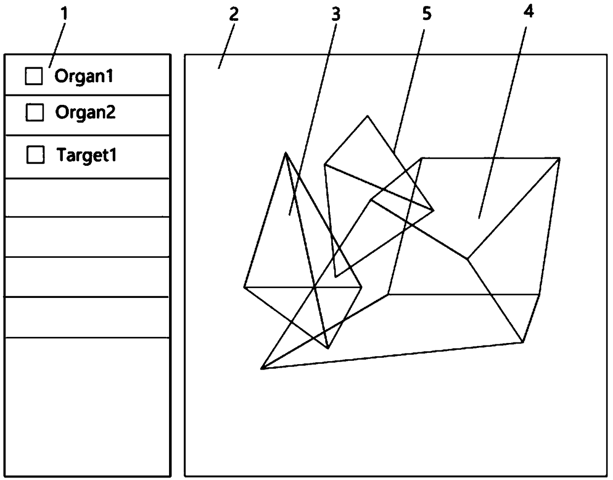
[0029] please participate Figure 1-2 As shown, the medical imaging image is obtained and the physician user outlines the tissue, organ and lesion data on the medical imaging image and is displayed in the image view area 2 after three-dimensional reconstruction, and the name of the tissue or lesion is displayed in the name list 1, by default , there are occlusion phenomena in tissues, organs and lesions in the 3D view, such as figure 1 The first tissue organ 3 and the second tissue organ 4 block the target organ 5 (or target lesion 5 ) to varying degrees. When the target organ 5 (or target lesion 5) is selected, the image display effect will be automatically adjusted in the image view area 2, as shown in figure 2 In the display, the occluded areas of the first tissue organ 3 and the second tissue organ 4 and the target organ 5 (or target lesion 5) are displayed in a fusion manner, and other areas are displayed in a blurred manner.


PUM


Abstract

The invention discloses a three-dimensional medical image fuzzy highlight display method, which relates to the technical field of tissue organ and lesion highlight in medical image. The method of theinvention comprises acquiring medical image images and outlining tissue organs and lesions; carrying out the three-dimensional reconstruction of target tissues and organs and target lesions to obtainthree-dimensional medical images; carrying out the fuzzy processing on the image region except the target lesion or the target tissue or organ in the medical three-dimensional image. Interacting the target lesion or the occlusion region of the target tissue or organ by adjusting the threshold value manually or adaptively; after the target lesion or target tissue is selected in the name list, restoring the original state of the medical 3D image. The method of the invention realizes the complete display of the target tissue organ and the target lesion in the medical three-dimensional image conveniently and quickly by carrying out the fuzzy processing on the image area except the target lesion or the target tissue organ on the medical three-dimensional image, and improves the accuracy rate ofmedical image analysis.

Description

technical field [0001] The invention belongs to the technical field of highlighting tissues, organs and lesions in medical images, and in particular relates to a fuzzy and highlighting display method for three-dimensional medical images. Background technique [0002] The existing 3D visualization system for medical imaging plays an important role in assisting doctors in diagnosis, surgery planning, radiotherapy planning and other fields. It can provide doctors with real and intuitive 3D images, which is convenient for doctors to observe and analyze from multiple angles, levels and aspects. Human organs and lesions. In real situations, human tissues, organs and lesions are mutually occluded. The existing technology highlights the target by adjusting the three-dimensional image lookup table (LUT) and hiding some organs and lesions. For example, in the radiotherapy planning system (TPS), the radiologist outlines the target area and organs at risk (organs at risk, OAR) for lung...

Claims


Application Information

Patent Timeline
Patent Type & Authority Applications(China)
IPC IPC(8): G06T19/00G06T19/20G06T17/00
CPCG06T17/00G06T19/003G06T19/20
Inventor 张庆林徐榭裴曦
Owner 安徽慧软科技有限公司
Why Eureka
Try Eureka