A Processing Method of Fundus Image Based on Neural Network

A neural network and processing method technology, applied in the field of fundus image processing, can solve the problems of low efficiency and low accuracy of diagnosis, and achieve the effect of improving efficiency and accuracy

Active Publication Date: 2022-02-01
CHANGZHOU IND TECH RES INST OF ZHEJIANG UNIV
View PDF7 Cites 0 Cited by
  • Summary
  • Abstract
  • Description
  • Claims
  • Application Information

AI Technical Summary

Problems solved by technology

The current judgment method is that the doctor makes a judgment by observing the fundus picture. Since the boundary between the optic cup area and the optic disc area in the fundus picture is not so clear, it takes a long time to judge the diameter ratio between them, and the diagnosis efficiency is low. less accurate

Method used


Image

  • A Processing Method of Fundus Image Based on Neural Network
  • A Processing Method of Fundus Image Based on Neural Network
  • A Processing Method of Fundus Image Based on Neural Network

Examples


Embodiment 1

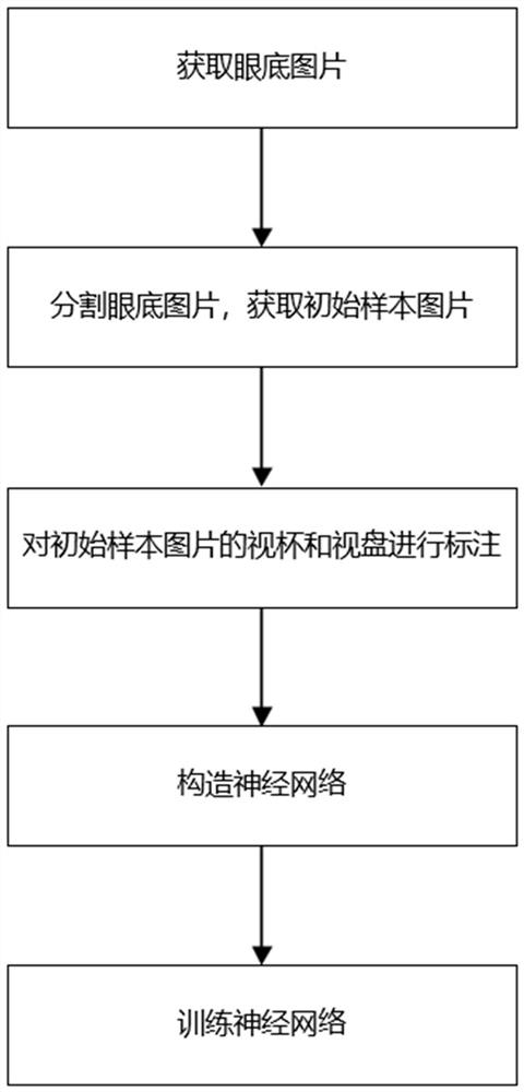
[0036] figure 1 Shown is a kind of embodiment 1 of the processing method of fundus picture based on neural network, comprises the following steps:

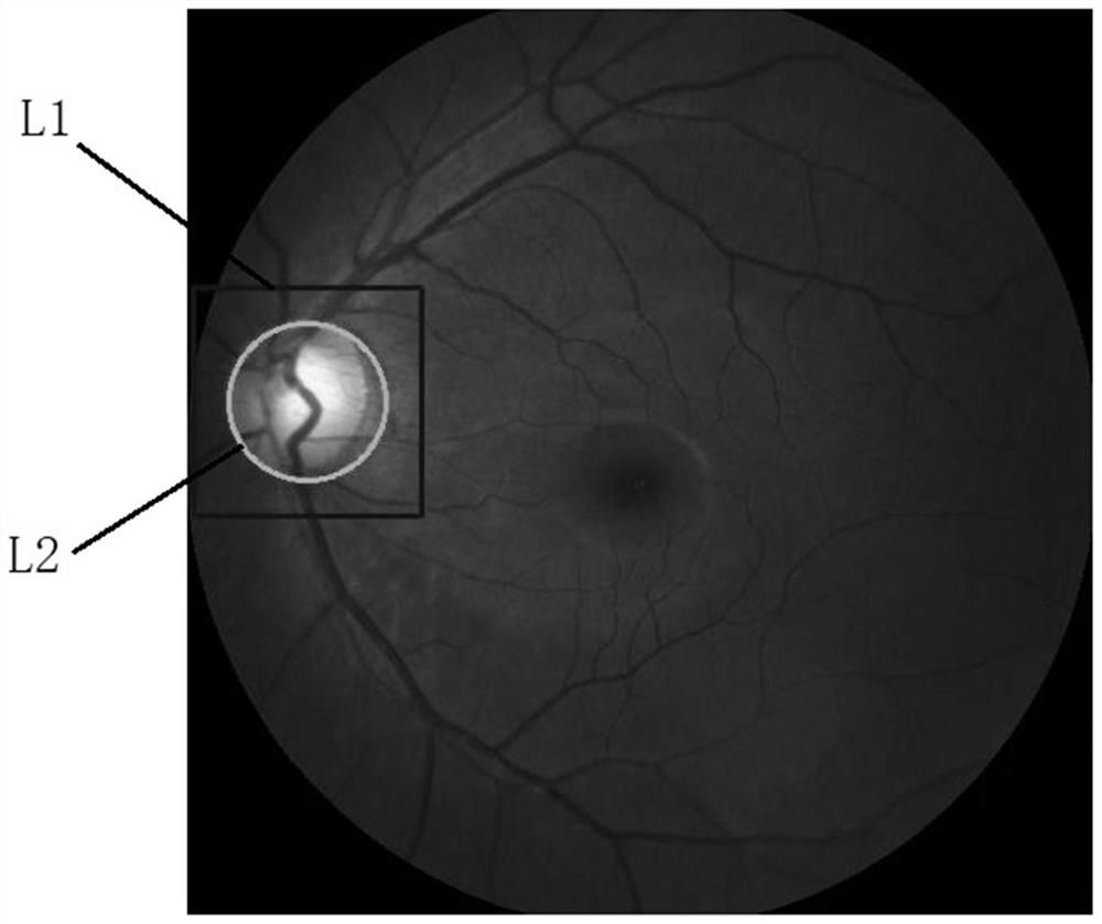
[0037] S1. Obtain several fundus pictures through camera shooting, figure 2 What is shown is a fundus picture. It can be seen that the actual picture taken by the fundus camera is relatively large, and most of the images used to identify and judge glaucoma come from the optic cup and disc. It is inconvenient to directly use the fundus picture to judge the cup and disc.

[0038] Therefore, in step S2, use the Hough transform circle detection method to find the circular area containing the optic disc; take the center of the circular area as the center, and use the preset length as the side length to cut out the optic disc and optic cup in the fundus image The square area of ​​is used as the initial sample image, from figure 2 It can be seen that the L1 part is a circular area, and the L2 part is a square area. Cutting out the o...


PUM


Abstract

The present invention provides a method for processing a fundus picture based on a neural network, comprising the following steps: S1, obtaining several fundus pictures through camera shooting; S2, detecting the region containing the optic disc and the optic cup in the fundus picture, and including The optic disc and optic cup areas are cut out from the fundus picture to obtain an initial sample picture; S3, circle the optic disc area and optic cup area on the initial sample picture and fill the optic disc area and optic cup area with different colors respectively to obtain training Sample pictures; S4, constructing a neural network; S5, training the neural network through several training sample pictures, and determining the parameters of the neural network; Efficient and accurate judgment improves the efficiency and accuracy of diagnosing glaucoma.

Description

technical field [0001] The invention relates to a processing method of fundus pictures, in particular to separating optic cups and optic discs from fundus pictures, and training a large number of samples to obtain a neural network for automatically identifying and segmenting optic cups and optic discs from fundus pictures. Background technique [0002] Glaucoma is one of the major eye diseases that lead to blindness. It is expected to affect about 80 million people by 2020. Unlike eye diseases such as cataracts and myopia, glaucoma vision loss is irreversible. Therefore, early screening is essential for early treatment It is essential to preserve vision and maintain quality of life. There are three clinical modalities used to screen for glaucoma: tonometry, visual field testing, and optic disc assessment. Intraocular pressure measurement has certain risks and is not enough to be an effective examination tool for a large number of glaucoma patients with normal intraocular pr...

Claims


Application Information

Patent Timeline
Patent Type & Authority Patents(China)
IPC IPC(8): G06T7/11G06T7/13G06N3/04
CPCG06T7/11G06T7/13G06T2207/30041G06N3/045
Inventor 覃鹏志包勇文耀锋
Owner CHANGZHOU IND TECH RES INST OF ZHEJIANG UNIV
Why Eureka
Try Eureka