
Method for automatically generating medical image diagnosis report based on deep learning method

A diagnostic report and medical imaging technology, applied in the field of radiology, can solve problems such as time-consuming, unsuitable for engineering, and limited practical effects, and achieve the effect of reducing workload and improving accuracy

Active Publication Date: 2018-12-21
HARBIN INST OF TECH +1
View PDF9 Cites 20 Cited by
  • Summary
  • Abstract
  • Description
  • Claims
  • Application Information

AI Technical Summary

Problems solved by technology

[0006] (1) For X-ray images without lesion labels, the accuracy rate is very limited
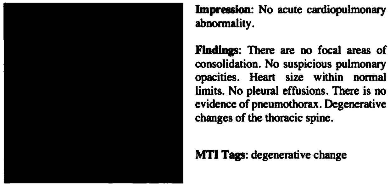
[0007] (2) The composition of text information is very cumbersome, and most diagnostic reports cannot have such complete information, and diagnostic reports with MTI Tags are even rarer
It takes a lot of time for doctors to summarize this kind of text information, which affects the efficiency of doctors' diagnosis
[0008] (3) For the unified calculation of loss for different models (CNN, sentence LSTM, word LSTM), different hyperparameters λ need to be set, which requires a lot of experiments and is not suitable for engineering
[0009] (4) At present, the most advanced and most used computer-aided diagnosis methods are CT images and PET image diagnosis. The existing technology is based on X-ray images, and the practical effect is very limited.

Method used


Image

  • Method for automatically generating medical image diagnosis report based on deep learning method
  • Method for automatically generating medical image diagnosis report based on deep learning method
  • Method for automatically generating medical image diagnosis report based on deep learning method

Examples


specific Embodiment approach 1

[0050] Specific embodiment 1: This embodiment provides a method for automatically generating a medical image diagnosis report based on a deep learning method. The specific implementation steps are as follows:

[0051] 1. Subject clustering of diagnosis reports based on LDA algorithm, and save diagnosis reports according to the subject. The medical imaging diagnosis report is a diagnosis report after text preprocessing, HMM Chinese word segmentation and skip-gram word embedding. Since each image corresponds to a segment of the diagnosis report, after subject clustering, each image will get the subject vector V corresponding to the diagnosis report. The dimension of the topic vector is the number of topics in the setting dimension, V i =1 means having topic i, V i =0 means there is no topic i.

[0052] 2. The position and size of the tumor in the default image of the system have been obtained, expressed by the center coordinate and radius. The subject vector is used as the label of...

specific Embodiment approach 2

[0056] Specific implementation manner 2: This implementation manner combines CT and PET image data to give a specific implementation process. The specific implementation process includes the following steps:

[0057] (1) Text preprocessing

[0058] Text preprocessing is to extract lung-related information in excel text, and remove some irrelevant characters. The text before processing is like image 3 Shown.

[0059] The overall implementation process of text preprocessing is as follows Figure 4 Shown. The main operation involves convenient reading of excel files. Use the python-based excel reading library xlrd to read in the file, remove the serial number, quotation marks, and punctuation before and after each line, and do keyword matching to extract text related to lung disease.

[0060] The processed text is like Figure 5 Shown.

[0061] (2) HMM Chinese word segmentation

[0062] Before training the HMM model, the text needs to be labeled with word segmentation, such as Image 6 S...


PUM


Abstract

The invention discloses a method for automatically generating a medical image diagnosis report based on the deep learning method. The method comprises steps that S1, a subject of the diagnosis reportis clustered based on the LDA algorithm, and the diagnosis report is saved separately according to the subject; S2, a subject vector is used as a label of each medical image; S3, CT images and PET images with different sizes are scaled to the same size as training data, subject vectors are used as labels, the VGGNet-19 is used as a network model for training, and a subject vector generation modelis obtained; S4, a text generation model is constructed; and S5, according to the subject vector of each image, the text of the corresponding subject is matched to obtain the diagnosis report of the image. The method is advantaged in that the method can be applied to scenes with images marked with lesions, a doctor has no need to manually summarize training data labels frequently, only the location and the size of the lesion should be marked, and the doctor's work is effectively reduced while the correct rate is improved.

Description

Technical field [0001] The invention belongs to the technical field of radiology, and relates to a method for generating a medical image diagnosis report, in particular to a method for automatically generating a medical image diagnosis report based on a deep learning method. Background technique [0002] In radiology, imaging physicians usually use CAD (Computer Aided Detection System) such as CT (Computer Tomography) and PET (Positron Emission Computed Tomography) to obtain patient image information. The image information is stored in a dicom format file. In addition to the main pixel information, the dicom file also contains a series of information such as the patient's name, gender, age, image type, and image serial number. The radiologist sums up the medical imaging information to obtain the examination findings, and obtains the patient's diagnosis opinion based on his own experience, and generates a diagnosis report. The diagnosis report includes the basic information of th...

Claims


Application Information

Patent Timeline
IPC IPC(8): G16H15/00G16H30/40G06F17/27
CPCG16H15/00G16H30/40G06F40/216G06F40/289
Inventor 苏统华于丽娟霍栋
Owner HARBIN INST OF TECH
Learn More