Breast cancer histopathologic grading method based on CNN and image histological feature fusion
A technology of radiomics and feature fusion, applied in the field of breast cancer histopathology grading, can solve the problem of inability to classify and distinguish, and achieve the effect of shortening the time of distinguishing and ensuring the accuracy of distinguishing
- Summary
- Abstract
- Description
- Claims
- Application Information
AI Technical Summary
Problems solved by technology
Method used
Image
Examples
Embodiment 1
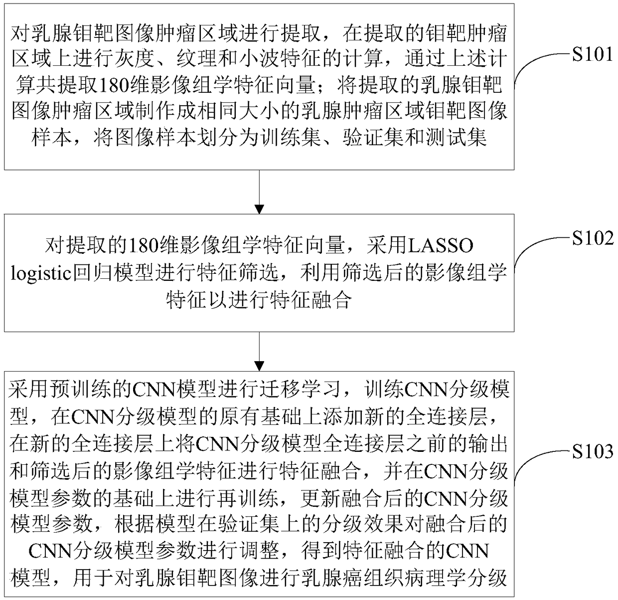
[0027] like figure 1 As shown, a method for grading breast cancer histopathology based on CNN and radiomics feature fusion of the present invention comprises the following steps:
[0028] Step S101: Extract the tumor area of the mammography image, and calculate grayscale, texture and wavelet features on the extracted mammography tumor area, and extract a total of 180-dimensional radiomics feature vectors through the above calculation; the extracted mammography The target image tumor area is made into mammary tumor image samples of the same size, and the image samples are divided into training set, verification set and test set.
[0029] Step S102: For the extracted 180-dimensional radiomics feature vector, the LASSO logistic regression model is used for feature screening, and the screened radiomics feature is used for feature fusion.
[0030] Step S103: Use the pre-trained CNN model for transfer learning, train the CNN classification model, add a new fully connected layer o...
Embodiment 2
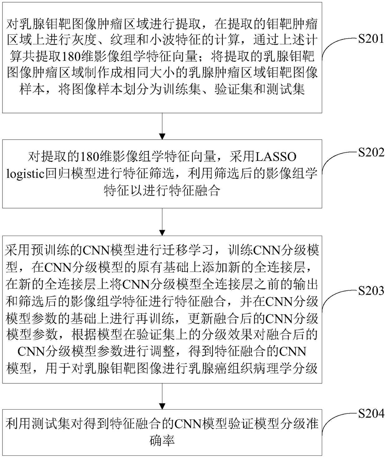
[0032] like figure 2 As shown, another breast cancer histopathological grading method based on CNN and radiomics feature fusion of the present invention comprises the following steps:
[0033] Step S201: Extract the tumor area of the mammography image, calculate the grayscale, texture and wavelet features on the extracted mammography tumor area, and extract a total of 180-dimensional radiomics feature vectors through the above calculation; the extracted mammography The target image tumor area is made into mammary tumor image samples of the same size, and the image samples are divided into training set, verification set and test set.
[0034] The step S201 includes:
[0035] Step S2011: extract the ROI from the tumor area of the mammography image to obtain the ROI image, calculate 14 grayscale features, 22 texture features and 144 wavelet features of the ROI image, and extract a total of 180-dimensional radiomics feature vectors;
[0036] The grayscale features are grays...
PUM
 Abstract
Description
Claims
Application Information
 - R&D
- Intellectual Property
- Life Sciences
- Materials
- Tech Scout
- Unparalleled Data Quality
- Higher Quality Content
- 60% Fewer Hallucinations
Browse by: Latest US Patents, China's latest patents, Technical Efficacy Thesaurus, Application Domain, Technology Topic, Popular Technical Reports.
© 2025 PatSnap. All rights reserved.Legal|Privacy policy|Modern Slavery Act Transparency Statement|Sitemap|About US| Contact US: help@patsnap.com


