Detection method of collagen terminal peptide
A detection method, collagen technology, applied in the field of collagen detection, can solve problems such as inapplicability, complexity, and false negatives, and achieve the effect of improving the range of detection capabilities
- Summary
- Abstract
- Description
- Claims
- Application Information
AI Technical Summary
Problems solved by technology
Method used
Image
Examples
Embodiment 1
[0041] Example 1 Detection of bovine type I collagen C-terminal antigen-specific band
[0042] One embodiment of the detection method of the collagen peptide telopeptide of the present invention comprises the following steps:
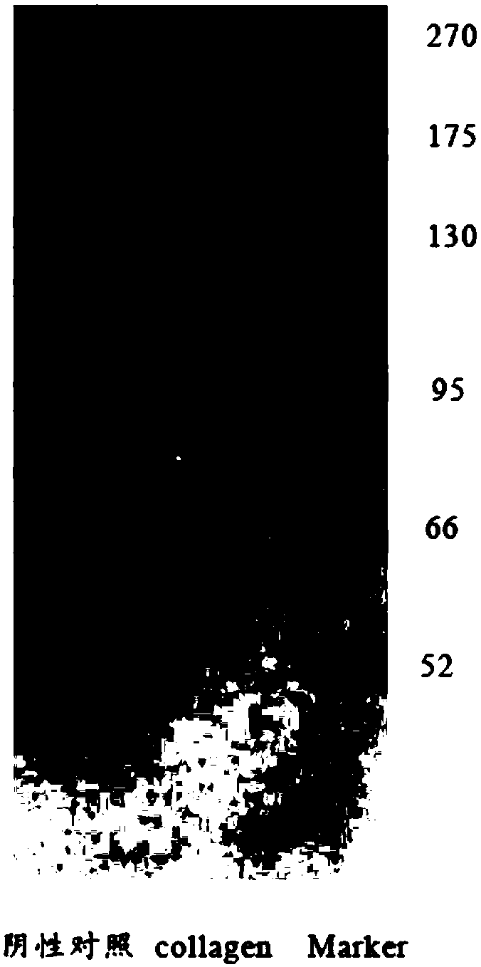
[0043] Sample processing: 5mL test sample (collagen containing telopeptide, such as figure 1 Collagen, collagen means collagen), 5mL negative control sample (collagen with telopeptide removed) were mixed with 5mL SDS buffer respectively, treated in boiling water bath at 100°C for 3min, cooled to room temperature, at 4°C, 5000rmp Centrifuge for 5 minutes to obtain the loading sample for later use; configure 6.5% separation gel and 4% stacking gel; the loading volume of each well is 10uL, and run at a current of 10mA for 30min. After the bromophenol blue in the sample enters the separation gel, adjust the electrophoresis The current is 20min.
[0044] Transfer: equilibrate the nitrocellulose membrane, 6 pieces of filter paper and the recovered gel in th...
Embodiment 2
[0048] Example 2 Excision effect test of bovine type I collagen C-terminal and N-terminal under different extraction time
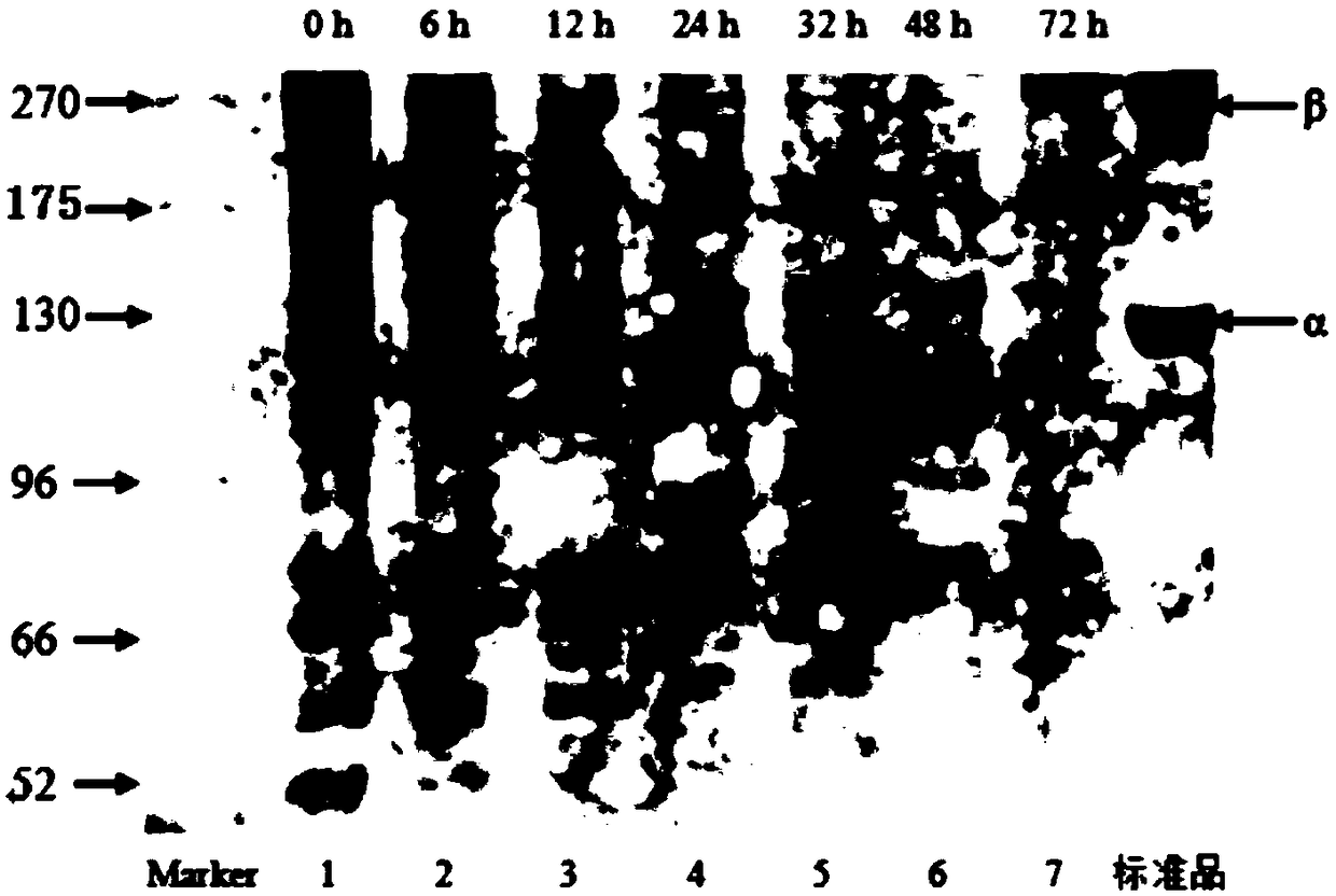
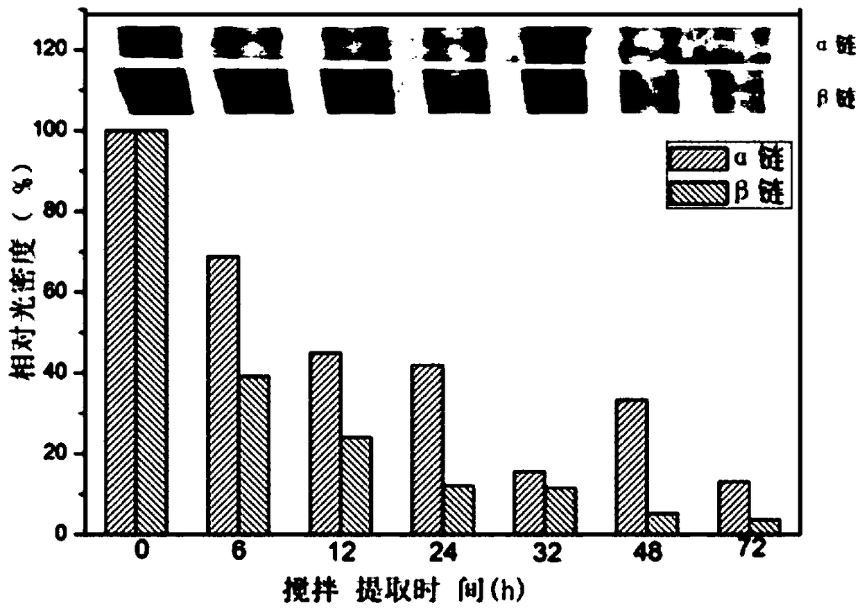
[0049] Sample description: Take about 200g of beef tendon, add 15g of pepsin, concentration of 0.5M, volume of 1L acetic acid solution, stir evenly, let it stand at 4°C for 14h, then place it in an electric mixer and stir at 4°C After extraction for 96 hours, the extracted collagen mixture was centrifuged at a speed of 10000rmp for 20 minutes at intervals, and used for western blot (ie westernblot) analysis.
[0050] One embodiment of the detection method of the collagen peptide telopeptide of the present invention comprises the following steps:
[0051] Sample processing: mix 5mL of the sample to be tested at the sampling time point with 5mL of SDS buffer, treat it in a boiling water bath at 100°C for 3min, cool to room temperature, and centrifuge at 5000rmp at 4°C for 5min to obtain the loaded sample for later use; configuration 6.5 % separation gel an...
Embodiment 3
[0058] Example 3 Detection of bovine type I collagen sponge N-terminal antigen-specific band
[0059] Sample description: Weigh three batches of collagen sponges of 0.05±0.0005g each, add 10mL sample buffer solution (100mM Tris, 8M urea, 2% SDS, 2% mercaptoethanol, pH8.0 ) mixing, at room temperature, shaking and dissolving for 4 hours, centrifuging at 5000rmp for 5 minutes, and taking the supernatant for later use.
[0060] One embodiment of the detection method of the collagen peptide telopeptide of the present invention comprises the following steps:
[0061] Sample processing: Mix 5mL of supernatant with 5mL of SDS buffer and treat it in a boiling water bath at 100°C for 3min. After cooling to room temperature, centrifuge at 4°C and 5000rmp for 5min to obtain the loading sample for later use; configure 6.5% separating gel and 4% stacking gel; the loading volume of each hole is 10uL, run at a current of 10mA for 30min, after the bromophenol blue in the sample enters the se...
PUM
 Abstract
Description
Claims
Application Information
 - Generate Ideas
- Intellectual Property
- Life Sciences
- Materials
- Tech Scout
- Unparalleled Data Quality
- Higher Quality Content
- 60% Fewer Hallucinations
Browse by: Latest US Patents, China's latest patents, Technical Efficacy Thesaurus, Application Domain, Technology Topic, Popular Technical Reports.
© 2025 PatSnap. All rights reserved.Legal|Privacy policy|Modern Slavery Act Transparency Statement|Sitemap|About US| Contact US: help@patsnap.com


