
A method for the isolation and bionic culture of pericytes in tumor tissue

A technology of tumor tissue and culture method, applied in the field of separation and bionic culture of pericytes in tumor cells, can solve problems such as difficulty in identification and no specific antibody labeling of pericytes

Active Publication Date: 2020-07-17
SUN YAT SEN MEMORIAL HOSPITAL SUN YAT SEN UNIV
View PDF1 Cites 0 Cited by
  • Summary
  • Abstract
  • Description
  • Claims
  • Application Information

AI Technical Summary

Problems solved by technology

[0012] In order to overcome the shortcomings and shortcomings of existing pericytes that are difficult to identify without specific antibody markers, the purpose of the present invention is to provide a method for identifying and isolating pericytes in tumor tissue through antibody labeling

Method used


Image

  • A method for the isolation and bionic culture of pericytes in tumor tissue
  • A method for the isolation and bionic culture of pericytes in tumor tissue
  • A method for the isolation and bionic culture of pericytes in tumor tissue

Examples


Embodiment 1

[0043] The present invention uses a new method to separate, purify and cultivate pericytes in tumor tissue. All separation operations are carried out in an ultra-clean bench, which includes the following steps in turn:

[0044] 1. Fresh tumor tissue samples are stored in normal saline containing 1% double antibody (v / v, penicillin and streptomycin) and 0.2% heparin (v / v), stored in an ice box, and within 4 hours Carry out enzymatic separation.

[0045] 2. Place the tissue in a 100mm petri dish, wash it with normal saline until it turns pale, and remove the coagulated blood with tweezers.

[0046] 3. Cut the tissue into pieces with sterilized scissors. Note: The more broken the cut, the better; do not use a homogenizer and razor blades, which will damage the cells; operate on ice.

[0047] 4. Enzyme hydrolysis solution: containing DMEM medium, 1% collagenase Type I, 1% collagenase Type III, 1% collagenase Type IV (Worthington) and 1% DNase (Roche, 100×). After enzymatic hydr...


PUM

PropertyMeasurementUnit
pore sizeaaaaaaaaaa
pore sizeaaaaaaaaaa
pore sizeaaaaaaaaaa

Abstract

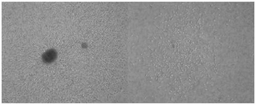
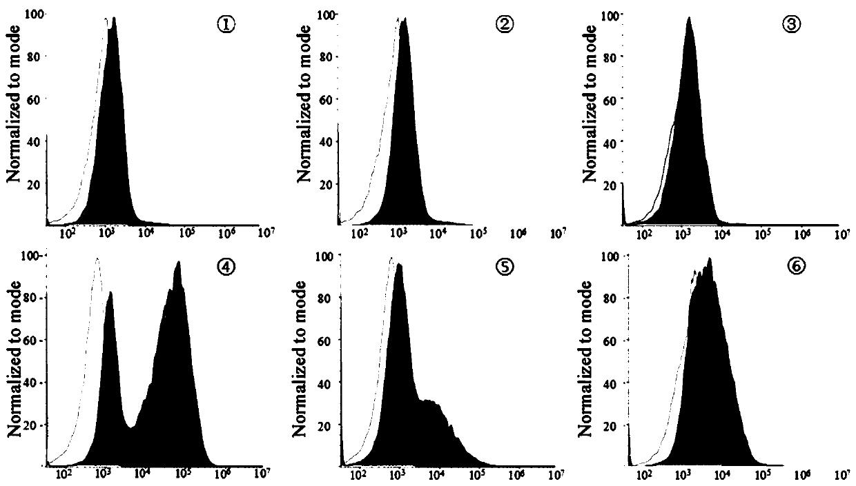
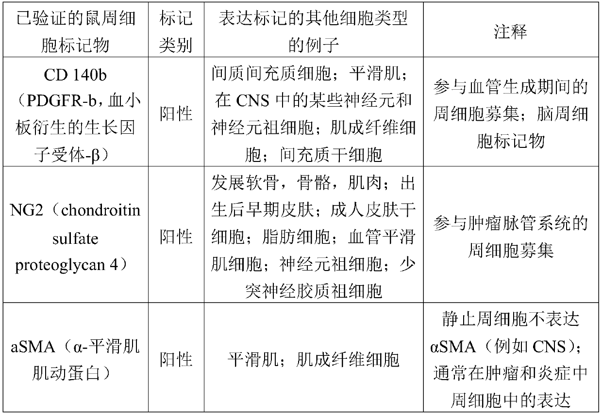
The invention discloses a method for separating and bionic culturing pericytes of tumor cells and relates to the field of metastasis of tumor. The method for separating and bionic culturing pericytesof tumor cells provides a departure from the traditional method of using two non-specific antibodies to label pericytes; 10 antibody labels including 5 positive labels and 5 negative labels are firstly used to separate, purify and identify pericytes. And through the morphological identification under a mircoscope, the pericytes can form microtubules in bionic culturing base, which can be seen obviously and which meet the morphological characteristics of pericytes, furthermore, the growth of pericytes in body is reproduced, and further explore of the function of the pericytes is based.

Description

technical field [0001] The invention relates to the field of tumor biology, in particular to a method for separating and bionically culturing pericytes in tumor cells. Background technique [0002] Pericytes are structural cells widely distributed throughout the microvascular walls throughout the body and, together with endothelial cells, form a barrier between microvessels and the interstitial space (1). Pericytes communicate with microvascular endothelial cells through cell junctions or paracrine signals, and play an important role in regulating microcirculatory blood flow, microvascular permeability and angiogenesis. Furthermore, in tumor tissues, pericytes have been shown to be functional cells that mediate endothelial maturation and stability and reduce vascular leakiness. Microvascular lesions in many diseases are accompanied by abnormal structure and function of pericytes, and the regulation of pericytes has become a research hotspot. Based on their morphology, peri...

Claims


Application Information

Patent Timeline
Patent Type & Authority Patents(China)
IPC IPC(8): C12N5/09G01N33/569
CPCC12N5/0693C12N2501/599C12N2509/00G01N33/56966
Inventor 黄炳培孟琼朱晓峰陈学曼孟亚明赵新保
Owner SUN YAT SEN MEMORIAL HOSPITAL SUN YAT SEN UNIV
Learn More