Biological three-dimensional printing preparation method of chondrocyte loading anti-inflammatory meniscus bracket
A chondrocyte and three-dimensional printing technology, applied in tissue regeneration, medical science, prosthesis, etc., to avoid the operation of in vitro cell culture and promote regeneration
- Summary
- Abstract
- Description
- Claims
- Application Information
AI Technical Summary
Problems solved by technology
Method used
Image


Examples
preparation example Construction
[0044] A method for preparing three-dimensional bioprinting of an anti-inflammatory meniscus stent loaded with chondrocytes. The method for preparing three-dimensional bioprinting of an anti-inflammatory meniscus stent loaded with chondrocytes includes the following steps:
[0045] S1. Construct a three-dimensional model of the meniscus:
[0046] 1. Data scanning: First, scan the knee joint on the healthy side of the patient with a three-dimensional scanning electron microscope, and then store the raw data obtained from the scan in the database;
[0047] 2. Data processing: segment and edit the knee joint model of the patient’s healthy side saved in step one through the bio-three-dimensional printer software to obtain a three-dimensional model of the outer or inner side of the knee joint meniscus. The three-dimensional model is mirrored to obtain a three-dimensional model of the meniscus suitable for the defect side of the patient, and then the three-dimensional meniscus model obtain...
Embodiment 1
[0073] 1. Perform nano-level 3D scanning electron microscopy on the left healthy knee joint of the patient, scan the original data, and store it in the database; segment and edit the knee joint image in the biological 3D printer software to obtain the outer side of the meniscus between the left knee joint Three-dimensional model. It is mirrored to obtain a three-dimensional model suitable for the outer side of the patient's right meniscus. Import the 3D data into the biological 3D printer software, perform partial correction and curved surface processing in turn, and save it as a stl format file.
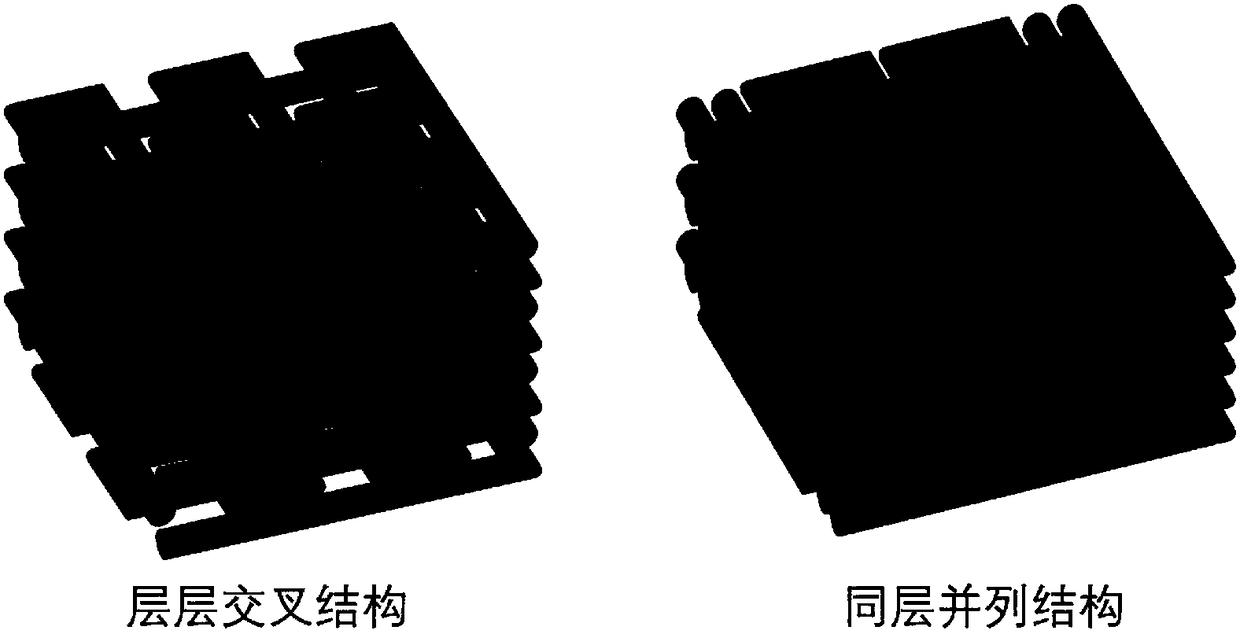
[0074] 2. The processed three-dimensional meniscus model is sliced and layered, and the printed polycaprolactone material and the hydrogel material loaded with fullerene and chondrocytes are respectively set into two printing paths. The two printing paths intersect layer by layer.
[0075] 3. After mixing the collagen and sodium alginate, add them to the DMEM medium, stir to dissolve...
Embodiment 2
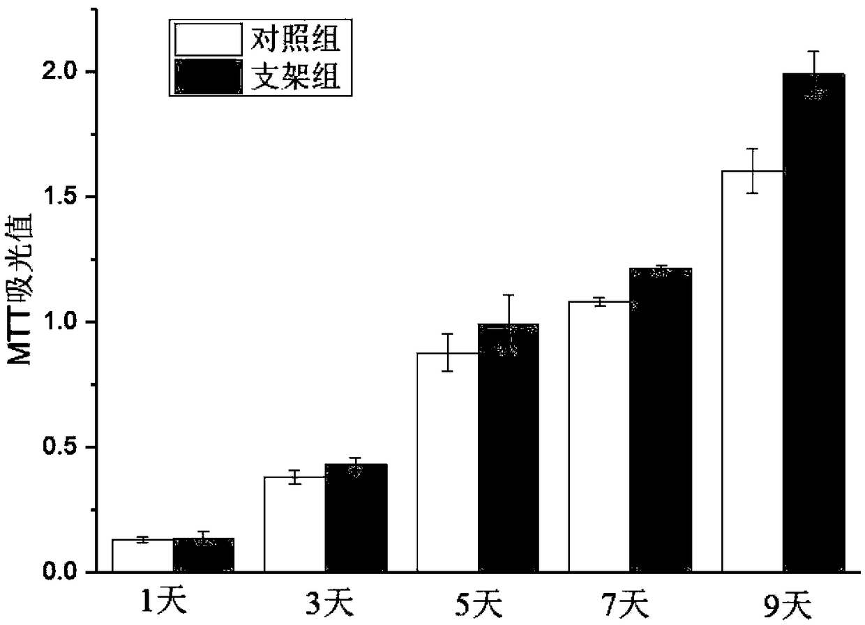
[0079] The chondrocyte-loaded anti-inflammatory meniscus scaffold prepared by using the three-dimensional bioprinting technology in Example 1 above was used to characterize the cell survival rate. The scaffold was placed in a 12-well plate for culture, and then the chondrocyte viability in the scaffold was detected by the MTT method. After culturing for 1, 3, 5, 7 and 9 days, discard the old medium and add PBS to rinse to remove dead cells. Add 500 μL of serum-free medium and 1500 μL of MTT solution to each well and incubate at 37°C for 4 hours. Then add 500μL of DMSO to each well to fully dissolve the formazan crystals, take 100μL of the dissolving solution and use a microplate reader to detect the absorbance at a wavelength of 492nm. Three parallel hole experiments were performed at each time point.
[0080] MTT results like image 3 It is shown that the chondrocytes on the scaffold have good proliferation behavior and their cell viability is good.
PUM
| Property | Measurement | Unit |
|---|---|---|
| diameter | aaaaa | aaaaa |
Abstract
Description
Claims
Application Information
 - Generate Ideas
- Intellectual Property
- Life Sciences
- Materials
- Tech Scout
- Unparalleled Data Quality
- Higher Quality Content
- 60% Fewer Hallucinations
Browse by: Latest US Patents, China's latest patents, Technical Efficacy Thesaurus, Application Domain, Technology Topic, Popular Technical Reports.
© 2025 PatSnap. All rights reserved.Legal|Privacy policy|Modern Slavery Act Transparency Statement|Sitemap|About US| Contact US: help@patsnap.com