
An apex detection device used in cardiac surgery

A surgical and cardiac technology, applied in surgery, surgical forceps, surgical lighting, etc., can solve the problems of difficult exploration of the apex of the heart, affect the surgical effect, and difficult to explore, and achieve the elimination of dark areas, uniform beam brightness, and avoid mutual infection. Effect

Active Publication Date: 2020-05-05
FUWAI HOSPITAL CHINESE ACAD OF MEDICAL SCI & PEKING UNION MEDICAL COLLEGE
View PDF8 Cites 0 Cited by
  • Summary
  • Abstract
  • Description
  • Claims
  • Application Information

AI Technical Summary

Problems solved by technology

[0002] Cardiac exploration is divided into extracardiac exploration and intracardiac exploration. The extracardiac exploration can be completed by the surgeon's fingers or tweezers and other instruments, while for the inner heart exploration, Due to the small incision and limited field of view, the exploration is difficult. General muscle retractors, tweezers, or suspension traction wires can only be effective for relatively shallow exposures, but it is difficult for the apex to be explored. Such problems are often encountered. For example, for a patient with ventricular septal dysplasia, the repair of multiple ventricular defects at the apex is the difficulty and focus of the entire operation. In order to completely distribute the apical defects, sometimes the right heart must be incised. This is a difficult problem. On the one hand, to accurately locate the apex of the heart, it is necessary to use right-angle forceps to repeatedly probe into the cavity of the right ventricle. The right coronary artery is avoided, so in order to take into account both internal and external aspects, it is necessary to repeatedly explore and move the young heart of the child during the operation. This is a great blow to the immature myocardium of the child and directly affects the effect of the operation.

Method used


Image

  • An apex detection device used in cardiac surgery
  • An apex detection device used in cardiac surgery
  • An apex detection device used in cardiac surgery

Examples


Embodiment Construction

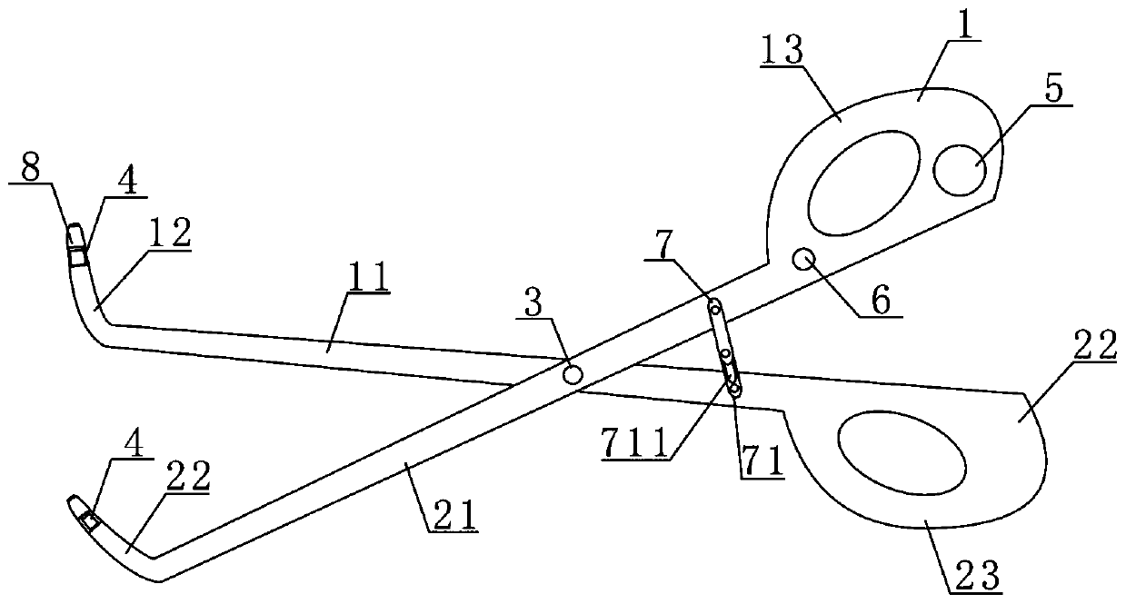
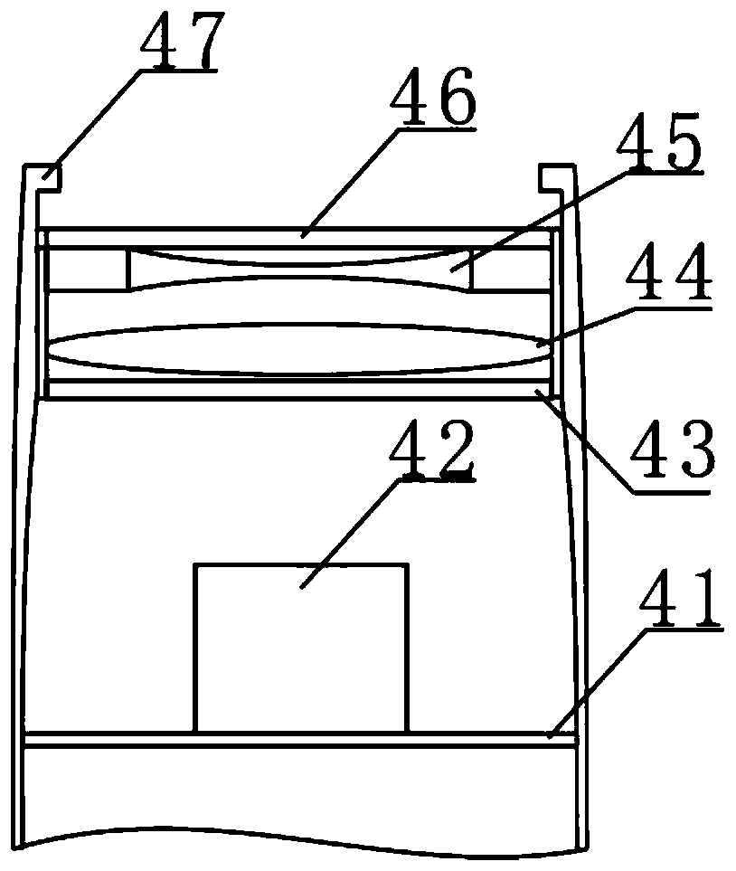
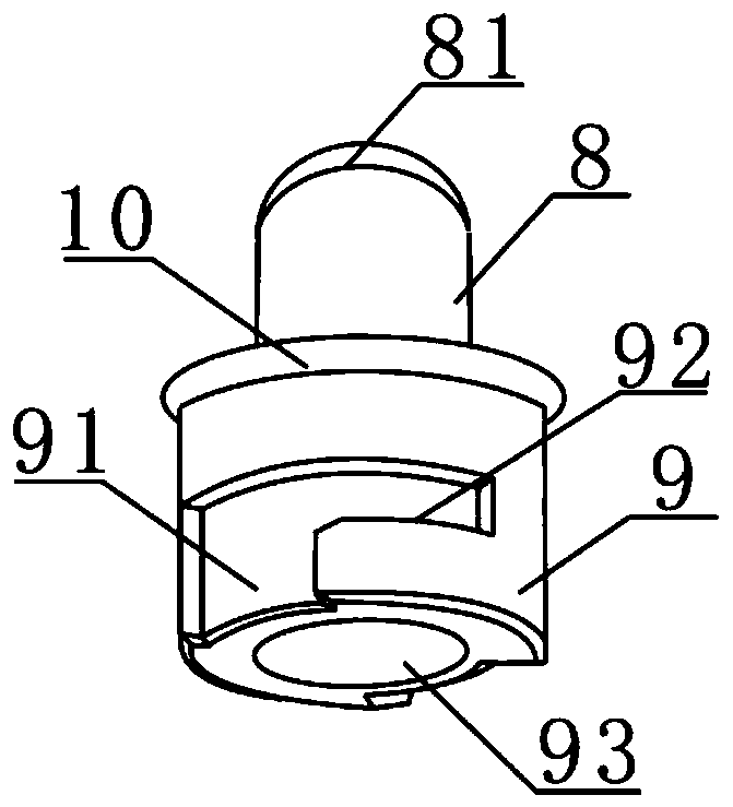
[0032] Such as figure 1 The shown apex detection device used in cardiac surgery includes a first clamp arm 1 and a second clamp arm 2 hinged together by a first fastener 3; the front end of the first clamp arm 1 is provided with The first elbow 11, the rear end of the first pincer arm 1 is provided with the first grip 12; the front end of the second pincer arm 2 is provided with the second elbow 21, the rear end of the second pincer arm The second grip part 22 is provided. The first elbow 11 and the second elbow 21 are hollow structures, and the front ends of the first elbow 11 and the second elbow 21 are respectively equipped with a laser light 42 detection assembly 4, the first elbow 11 and the second elbow The front ends of the two elbows 21 are respectively provided with a light guide column 8 detachably connected thereto.

[0033] Wherein, the light guide column 8 includes a light guide body, the rear end of the light guide body is provided with a connector 9 detachably...


PUM


Abstract

The invention discloses a heart tip exploring device used in a cardiac surgical operation. The heart tip exploring device comprises a first forceps arm and a second forceps arm which are hinged together through a first fastener. A first bend is arranged at the front end of the first forceps arm, and a first held portion is arranged at the rear end of the first forceps arm. A second bend is arranged at the front end of the second forceps arm, and a second held portion is arranged at the rear end of the second forceps arm. The first bend and the second bend are of hollow structures, a laser lamp exploring assembly is arranged in the front end of the first bend and the front end of the second bend respectively, and a light guiding column detachably connected with the front ends of the bends are arranged at the front end of the first bend and the front end of the second bend respectively. Each light guiding column comprises a light guiding body, wherein a connector detachably connected with the first bend and the second bend is arranged at the rear end of the light guiding body, a light scattering face is arranged at the front end of the light guiding column, and an outward protruding bag is arranged at the connecting position of the light guiding column and the connector.

Description

technical field [0001] The present invention relates to an apex detection device used in cardiac surgery. Background technique [0002] Cardiac exploration is divided into extra-cardiac exploration and intra-cardiac exploration. The extra-cardiac exploration can be completed with the surgeon's fingers or tweezers and other instruments. For the inner heart exploration, due to the small incision and limited field of view, it is more difficult to explore. Retractors, tweezers, or suspension traction wires can only be effective for relatively shallow exposures, but it is difficult to detect the apex of the heart. The inventors often encounter such problems during cardiac surgery, such as for a patient with ventricular septal dysplasia. For patients, the repair of multiple ventricular defects at the apex is the difficulty and focus of the entire operation. In order to completely distribute the apical defects, sometimes the right heart needs to be incised. This is a difficult prob...

Claims


Application Information

Patent Timeline
Patent Type & Authority Patents(China)
IPC IPC(8): A61B17/28A61B90/13A61B17/12A61B90/30
CPCA61B17/12A61B17/282A61B17/2833A61B17/2841A61B90/13A61B90/30A61B2017/12004
Inventor 彭博王强
Owner FUWAI HOSPITAL CHINESE ACAD OF MEDICAL SCI & PEKING UNION MEDICAL COLLEGE
Learn More