
Endoscope operating sheath for urological surgery

A technique for urology and endoscopy, applied to the field of endoscopy, can solve the problems of increasing the cost of surgery, increasing the difficulty of surgery, and unsmooth surgery, and achieves the effect of simple method and easy operation.

Pending Publication Date: 2017-11-07
YOUCARE TECH CO LTD
View PDF4 Cites 15 Cited by
  • Summary
  • Abstract
  • Description
  • Claims
  • Application Information

AI Technical Summary

Problems solved by technology

Clinical internal inspection and visualized surgical treatment require endoscopes with clear images to help doctors improve the accuracy of diagnosis, but the existing endoscopes used for flexible ureteroscopic surgery are flexible endoscopes, although they will not The human body is damaged during the operation, but because the hose is difficult to move forward after encountering resistance, it is difficult for the hose to accurately reach the surgical site, and it cannot accurately and completely reflect the lesion of the lesion, resulting in an unsmooth operation process, increasing the difficulty of the operation, and increasing the operation time. time and patient pain, and also raises the cost of surgery

Method used


Image

  • Endoscope operating sheath for urological surgery
  • Endoscope operating sheath for urological surgery
  • Endoscope operating sheath for urological surgery

Examples


Embodiment Construction

[0042] The present invention will be further described in detail below in conjunction with the accompanying drawings and specific embodiments.

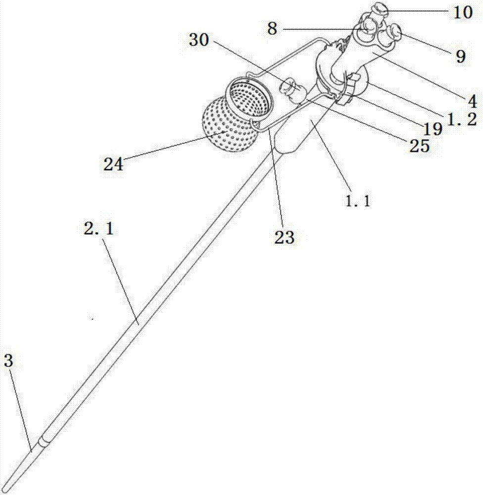
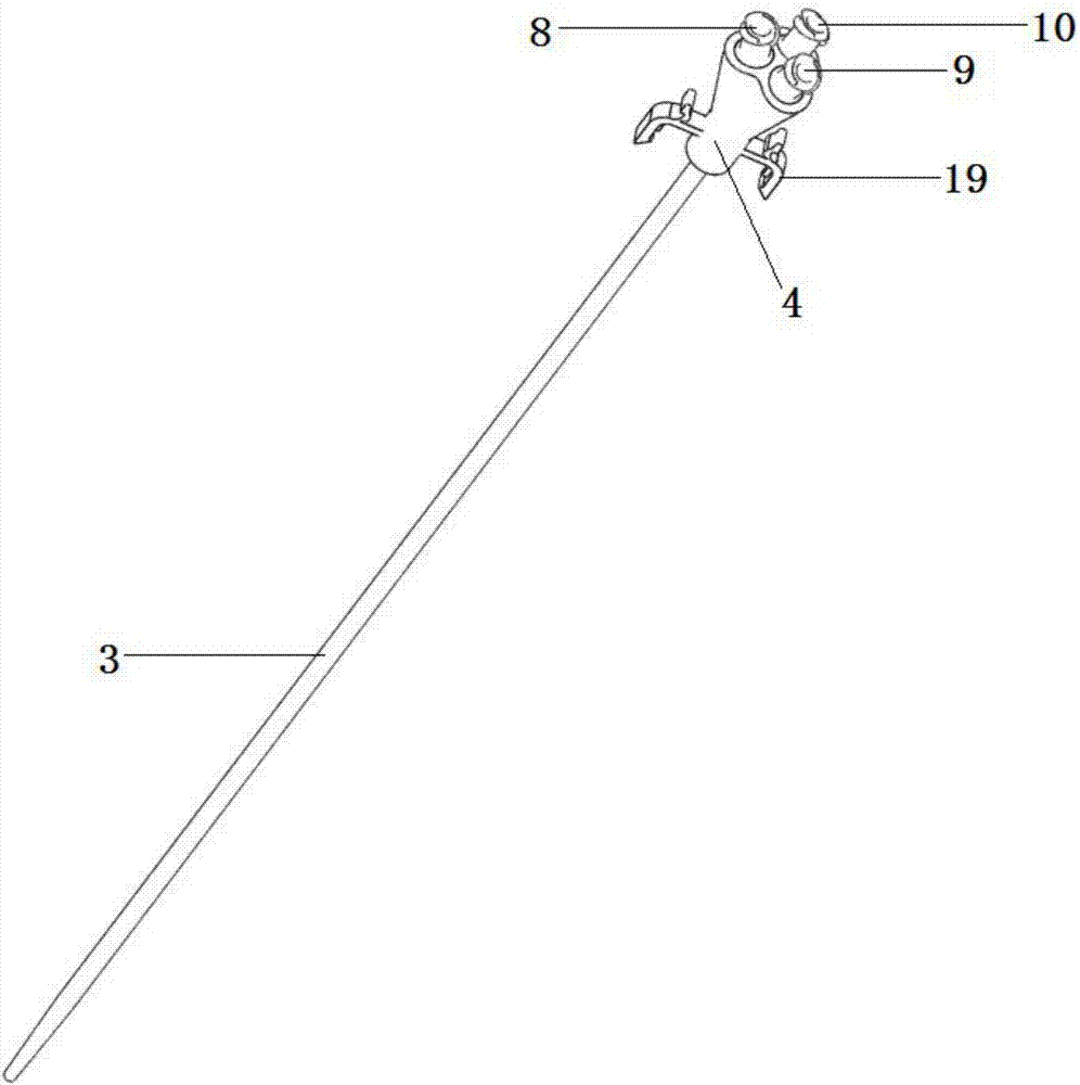
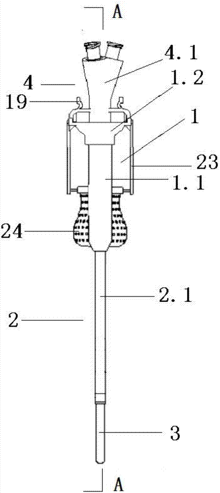
[0043] Such as figure 1 The endoscope working sheath for urological surgery shown in -18 includes an endoscope handle 1, an endoscope working sheath 2 and can be arranged in the endoscope working sheath 2, opposite to the endoscope working sheath 2 Sliding mandrel3. Mandrel 3 is made of medical polymer material (but not limited to polymer material), which has a certain degree of plasticity and can be deformed and bent to a certain extent, so that it can enter the human body and reach the diseased part more conveniently.

[0044] Endoscope working sheath 2 can be hard sheath 2.1 or soft and hard sheath (including hard sheath 2.1 and soft sheath 2.2):

[0045] Such as figure 1 —6 and Figure 17 As shown, the rear end of the hard sheath 2.1 is fixed in the endoscope handle 1, and the front end is located outside the endoscope handle ...


PUM


Abstract

The invention discloses an endoscope operating sheath for urological surgery. The endoscope operating sheath comprises a core rod which can be arranged in the endoscope operating sheath to axially slide relative to the endoscope operating sheath. The core rod is internally provided with a guide wire passage, a water injection passage and an image passage, and the guide wire passage, the water injection passage and the image passage extend from the rear end to the front end of the core rod. An endoscope handle is provided with a guide wire connector, a water injection connector and an endoscope connector for guiding a guide wire, water and an endoscope into the guide wire passage, the water injection passage and the image passage respectively. The endoscope operating sheath and the core rod enter a human body along the guide wire, the core rod is drawn out after reaching a lesion part, then a flexible cystoscope is put in the endoscope operating sheath, and the endoscope operating sheath and the flexible cystoscope are operated to observe the lesion part and cooperate with surgical instruments for performing the surgery. The method is simple and easy to operate, and the endoscope operating sheath can be widely applied to renal pelvis and renal caliceal calculi surgery to serve as a visible ureterectasia sheath and is applicable but not limited to cardiovascular intervention, gynecology department, thyroid gland and mammary gland department, neurosurgery department and orthopedics department and other unmentioned related clinical medical fields.

Description

technical field [0001] The invention relates to the technical field of endoscopes, in particular to an endoscope working sheath structure for urological surgery. Background technique [0002] Endoscope is a high-end medical device that integrates material science, traditional optics, ergonomics, precision machinery, modern electronics, mathematics, and software. It is widely used in medical treatment. In flexible ureteroscopic surgery, the flexible ureteroscope can enter the body through the urethra to observe the lesion and can cooperate with surgical instruments to perform visual surgery in the body. Clinical internal inspection and visualized surgical treatment require endoscopes with clear images to help doctors improve the accuracy of diagnosis, but the existing endoscopes used for flexible ureteroscopic surgery are flexible endoscopes, although they will not The human body is damaged during the operation, but because the hose is difficult to move forward after encount...

Claims


Application Information

Patent Timeline
Patent Type & Authority Applications(China)
IPC IPC(8): A61B17/00A61B17/22
CPCA61B17/00234A61B17/22A61B2017/00831A61B2017/0034A61B2017/00336A61B2017/00296A61B2017/00238A61B2017/22072A61B2017/22038A61B2017/22082A61B2017/22079
Inventor 龙刚李金平毛业云格伦·迈克尔·普雷明格叶章群李建兴余虓张军晖肖河李文成胡学成吴耀辉
Owner YOUCARE TECH CO LTD
Learn More