
Magnetic resonance image processing method and device

A magnetic resonance image and processing method technology, applied in the field of magnetic resonance image processing methods and devices, can solve the problems of inaccurate ischemic penumbra areas, inability to truly reflect the ischemia and bleeding conditions of brain tissue, and complicated operations, etc. Achieve the effect of saving image processing process, reducing complexity and improving security

Active Publication Date: 2017-09-05
SIEMENS HEALTHINEERS LTD +2
View PDF4 Cites 8 Cited by
  • Summary
  • Abstract
  • Description
  • Claims
  • Application Information

AI Technical Summary

Problems solved by technology

[0005] However, because the diffusion model of DWI cannot truly reflect the ischemia and hemorrhage of brain tissue, the ischemic penumbra area determined by the prior art is not accurate
[0006] Also, PWI scans usually require the injection of contrast fluid, so the operation is complicated and has safety issues

Method used


Image

  • Magnetic resonance image processing method and device
  • Magnetic resonance image processing method and device
  • Magnetic resonance image processing method and device

Examples


Embodiment Construction

[0053] In order to make the technical solutions and advantages of the present invention clearer, the present invention will be further described in detail below with reference to the accompanying drawings and embodiments. It should be understood that the specific embodiments described herein are only used to illustrate the present invention, and are not used to limit the protection scope of the present invention.

[0054] For the sake of brevity and intuition in description, the solution of the present invention is explained below by describing several representative embodiments. Numerous details in the embodiments are provided only to aid in understanding the aspects of the invention. However, it is obvious that the technical solutions of the present invention may not be limited to these details during implementation. In order to avoid unnecessarily obscuring aspects of the present invention, some embodiments are not described in detail, but merely framed. Hereinafter, "inc...


PUM


Abstract

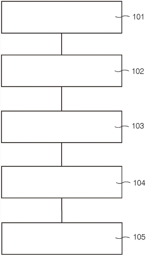
The embodiment of the invention discloses a magnetic resonance image processing method and device. The method comprises the steps that diffusion weighted imaging scanning and pulse parameter modulation imaging scanning based on chemical exchange saturation transfer are performed on the area of interest of the brain, and a diffusion weighted image and a chemical exchange saturation transfer image are acquired; when the chemical exchange saturation transfer image presents low signal shadow in comparison with the preset benchmark template, arterial spin mark imaging scanning is performed on the area of interest of the brain so as to acquire an arterial spin mark image; the diffusion weighted image is compared with the arterial spin mark image so as to determine a first mismatch area; the diffusion weighted image is compared with the chemical exchange saturation transfer image so as to determine a second mismatch area; and the second mismatch area is removed out of the first mismatch area, and the residual area of the first mismatch area is determined as an ischemic penumbra area. The accuracy of the ischemic penumbra can be enhanced, the image processing flow can be saved, the complexity can be reduced and the safety can be enhanced.

Description

technical field [0001] The present invention relates to the technical field of magnetic resonance imaging, in particular to a magnetic resonance image processing method and device. Background technique [0002] Magnetic resonance imaging (Magnetic Resonance Imaging, MRI) is a biomagnetic nuclear spin imaging technology that has developed rapidly with the development of computer technology, electronic circuit technology, and superconductor technology. It uses magnetic fields and radio frequency pulses to vibrate the hydrogen nuclei (ie H+) in human tissue to generate radio frequency signals, which are processed and imaged by a computer. When an object is placed in a magnetic field, irradiated with appropriate electromagnetic waves to make it resonate, and then the electromagnetic waves released by it are analyzed, the position and type of the nuclei that make up the object can be known. Stereoscopic image. For example, an animation of a serial slice that can be obtained by ...

Claims


Application Information

Patent Timeline
IPC IPC(8): G06T7/11
CPCG06T2207/10088G06T2207/30016
Inventor 王梅云龚启勇白岩钱天翼马潇越刘太元
Owner SIEMENS HEALTHINEERS LTD
Learn More