
RNA-protein-DNA in-situ multiple staining method of tissue chip

A tissue chip, -DNA technology, applied in the field of molecular biology, can solve the problem of not being able to detect and obtain biological information, and achieve the effect of reducing costs

Inactive Publication Date: 2017-06-20
SHENZHEN UNIV +1
View PDF3 Cites 3 Cited by
  • Summary
  • Abstract
  • Description
  • Claims
  • Application Information

AI Technical Summary

Problems solved by technology

[0007] The purpose of the present invention is to provide a RNA-protein-DNA in situ multiple staining method for tissue chips, aiming at solving the problem of inability to detect three molecular levels of RNA, protein and DNA on the same tissue chip in the prior art and obtain corresponding bioinformatic questions

Method used


Image

  • RNA-protein-DNA in-situ multiple staining method of tissue chip
  • RNA-protein-DNA in-situ multiple staining method of tissue chip
  • RNA-protein-DNA in-situ multiple staining method of tissue chip

Examples


no. 1 example

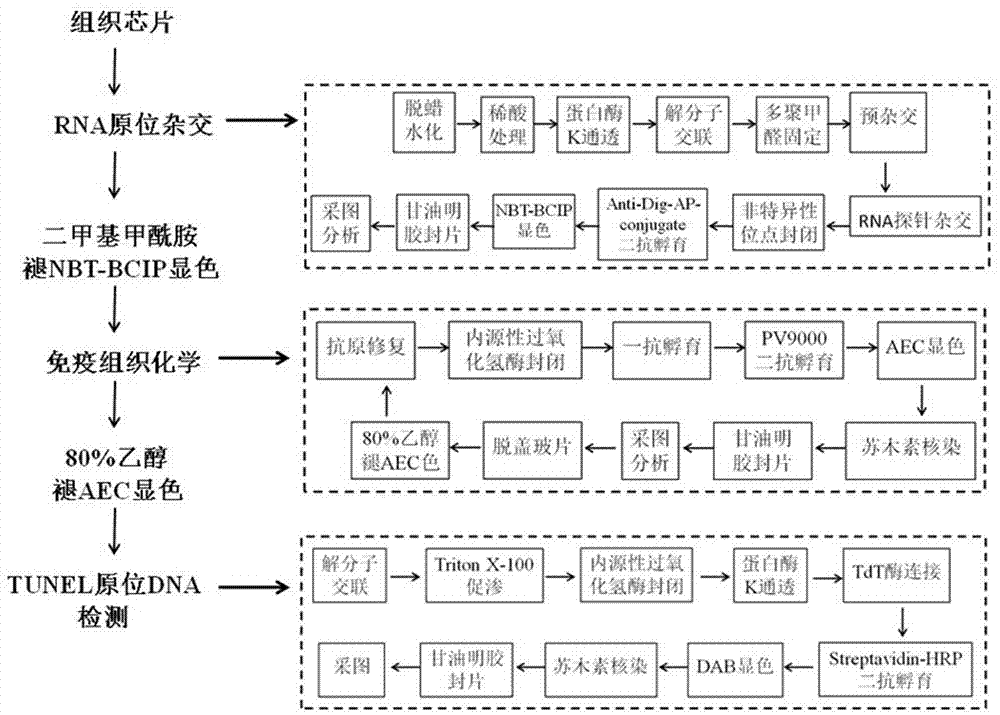
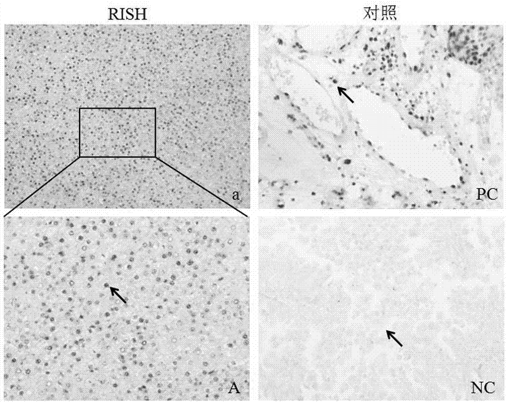
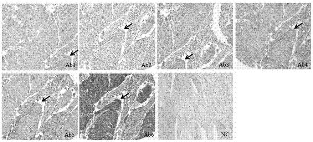
[0038] The present invention proposes a first embodiment, an RNA-protein-DNA in situ multiple staining method for a tissue chip, the method sequentially performs RNA in situ hybridization, immunohistochemical staining and DNA in situ hybridization on the same tissue chip;

[0039] In the process of RNA in situ hybridization, immunohistochemical staining and DNA in situ hybridization, an epitope antigen repair steamer is used to perform molecular cross-linking and unzipping or antigen repair on the tissue chip;

[0040] During RNA in situ hybridization and immunohistochemical staining, the slides were sealed with gelatin and glycerin after color development.

[0041] It should be noted that the epitope antigen retrieval steamer can effectively repair the "molecular mask" formed in the tissue section on the tissue chip fixed by paraformaldehyde, fully expose the antigenic determinant, and cross-link and dissolve with other molecules or Compared with antigen retrieval methods (su...


PUM


Abstract

The invention relates to the field of molecular biology, and provides a RNA-protein-DNA in-situ multiple staining method of a tissue chip. The method comprises the following steps: subjecting a tissue chip to RNA in-situ hybridization, immumohistochemical staining, and DNA in-situ hybridization in sequence; during the processes of RNA in-situ hybridization, immumohistochemical staining, and DNA in-situ hybridization, subjecting the tissue chip to molecule crosslinking melting or antigen retrieval by adopting an epitope antigen retrieving steamer; and during the processes of RNA in-situ hybridization and immumohistochemical staining, sealing the chip by gelatin glycerol after color development. The provided method can detect one tissue chip in three molecular levels (RNA, protein, and DNA), can obtain more comprehensive biological information, and reduces the cost of obtaining the biological information.

Description

technical field [0001] The invention relates to the field of molecular biology, in particular to an RNA-protein-DNA in situ multiple staining method of a tissue chip. Background technique [0002] Classical immunohistochemistry refers to the use of a known primary antibody (primary antibody) to specifically bind to the targeted target antigen on a single tissue section, and then the use of an enzyme-labeled secondary antibody (secondary antibody) to bind to the primary antibody. The multivalent binding of anti-antibody and the color development of enzyme substrates are used to amplify the immune reaction, so that the distribution and expression of the tested antigen / protein in tissue cells can be clearly observed under the microscope. This method has become one of the most important and influential experimental diagnostic techniques widely used in the fields of basic medicine and clinical medicine. [0003] In the past two decades, with the revolutionary breakthrough of ant...

Claims


Application Information

Patent Timeline
IPC IPC(8): G01N1/30G01N21/78
Inventor 盛司潼王筠邓芳蒋春樊游绍进
Owner SHENZHEN UNIV
Learn More