
Method of repairing human articular cartilage based on 3D bioprinting

A bioprinting, articular cartilage technology, applied in tissue regeneration, medical science, prosthesis, etc., can solve the problems of cartilage degeneration tissue transplantation, cartilage necrosis, failure and other problems, achieve low cytotoxicity, simple and fast operation, and accurate operation. Effect

Inactive Publication Date: 2017-02-22
武汉枫霖科技有限公司
View PDF0 Cites 4 Cited by
  • Summary
  • Abstract
  • Description
  • Claims
  • Application Information

AI Technical Summary

Problems solved by technology

This step actually causes additional necrosis of the existing cartilage and eventually leads to cartilage degeneration and tissue graft failure

Method used


Image

  • Method of repairing human articular cartilage based on 3D bioprinting
  • Method of repairing human articular cartilage based on 3D bioprinting
  • Method of repairing human articular cartilage based on 3D bioprinting

Examples


Embodiment Construction

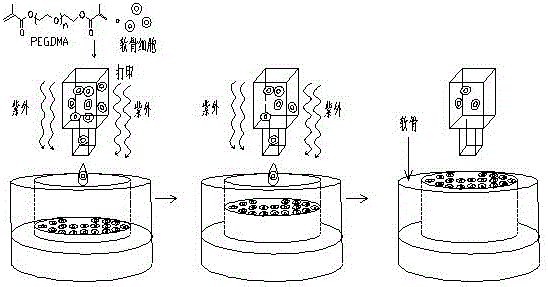
[0025] like figure 1 As shown, the present invention discloses a method for repairing human articular cartilage based on 3D bioprinting, comprising the following steps:

[0026] Preparation of PEGDMA

[0027] 3 kDa PEG (polyethylene glycol) was dissolved in tetrahydrofuran and PEG was reacted with methacryloyl chloride overnight under nitrogen. The synthesized PEGDMA (polyethylene glycol dimethacrylate) macromonomer was purified by ether precipitation and overnight freeze-drying, and detected by proton nuclear magnetic resonance to ensure that the purity of the synthesized PEGDMA macromonomer was greater than 95%.

[0028] Human mesenchymal stem cell isolation

[0029] Take the healthy adult bone marrow fluid under sterile conditions, add an equal volume of PBS, and disperse the cells by pipetting. The cells were collected by centrifuging 300 g of bone marrow PBS mixture for 5 min at room temperature. Cells were resuspended in PBS and counted. Take 300 g of bone marrow PB...


PUM

PropertyMeasurementUnit
Strengthaaaaaaaaaa

Abstract

The invention relates to a method for repairing human articular cartilage based on 3D bioprinting. The method comprises the following steps: a) PEGDMA is prepared from PEG (polyethylene glycol)and methacryloyl chloride and purified; b) human mesenchymal stem cells are separated; c) the human mesenchymal stem cells are subjected to passage and adjusted in density, an induction medium is replaced for culture after cell attachment; d) a bovine femur is washed, a full-thickness cartilage defect is formed at the center of an osteochondral embolism by a sterile living body perforator, and the osteochondral embolism is placed in a DMEM (dulbecco's modified eagle medium) and cultivated into 3D biopaper; e) the purified PEGDMA is dissolved, a photoinitiator 2959 is added, and chondrocytes obtained through induction in the step c) are re-suspended in a filtered and sterilized PEGDMA solution for preparation of bio-ink; f) the bio-ink is added to a disinfected and sterilized bioprinter, and the 3D cartilage is printed on the 3D biopaper according to the designed shape and size and is cultured in the induction medium. The method has the advantages of being simple and quick to operate, accurate, low in cytotoxicity and is expected to become a clinical cartilage repair method.

Description

technical field [0001] The invention relates to the technical field of tissue engineering, in particular to a method for repairing human articular cartilage based on 3D bioprinting. Background technique [0002] Cartilage defects due to osteoarthritis, aging, and joint damage are among the leading causes of joint pain and chronic disability. Mature cartilage cannot heal on its own due to the lack of blood vessels, nerves and lymph. Currently the most commonly used treatment for advanced cartilage degeneration is joint replacement, but the operation itself is complicated, often causes surrounding tissue infection, and is very expensive. Although tissue engineering therapies based on cell transplantation were introduced more than two decades ago, current cartilage tissue engineering strategies have not been able to create the same tissue as native cartilage, including hierarchical structure, extracellular matrix, and mechanical properties. In addition, almost all knee cartil...

Claims


Application Information

Patent Timeline
IPC IPC(8): A61L27/38A61L27/36A61L27/18B33Y70/00
CPCA61L27/3817A61L27/18A61L27/3612A61L27/3654A61L27/3852A61L2430/06B33Y70/00
Inventor 崔晓峰高桂芳
Owner 武汉枫霖科技有限公司
Learn More