
Method and apparatus for improved endothelial implantation

A technology of endothelial and injection devices, applied in eye implants, parts of surgical instruments, medical science, etc., can solve problems such as damage

Inactive Publication Date: 2017-02-15
E K - D D S
View PDF3 Cites 0 Cited by
  • Summary
  • Abstract
  • Description
  • Claims
  • Application Information

AI Technical Summary

Problems solved by technology

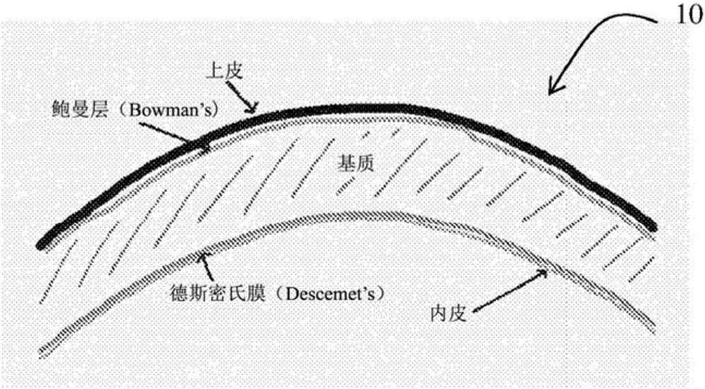
The handle extending through the ring has been found to risk contacting the De Smith's membrane to be grafted and risking damaging it

Method used


Image

  • Method and apparatus for improved endothelial implantation
  • Method and apparatus for improved endothelial implantation
  • Method and apparatus for improved endothelial implantation

Examples


Embodiment Construction

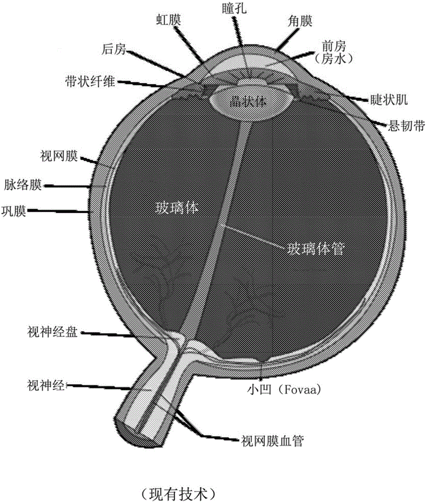
[0077] figure 1 Is a schematic diagram of the eyeball showing the iris, pupil, cornea, lens, vitreous, optic nerve, and other parts.

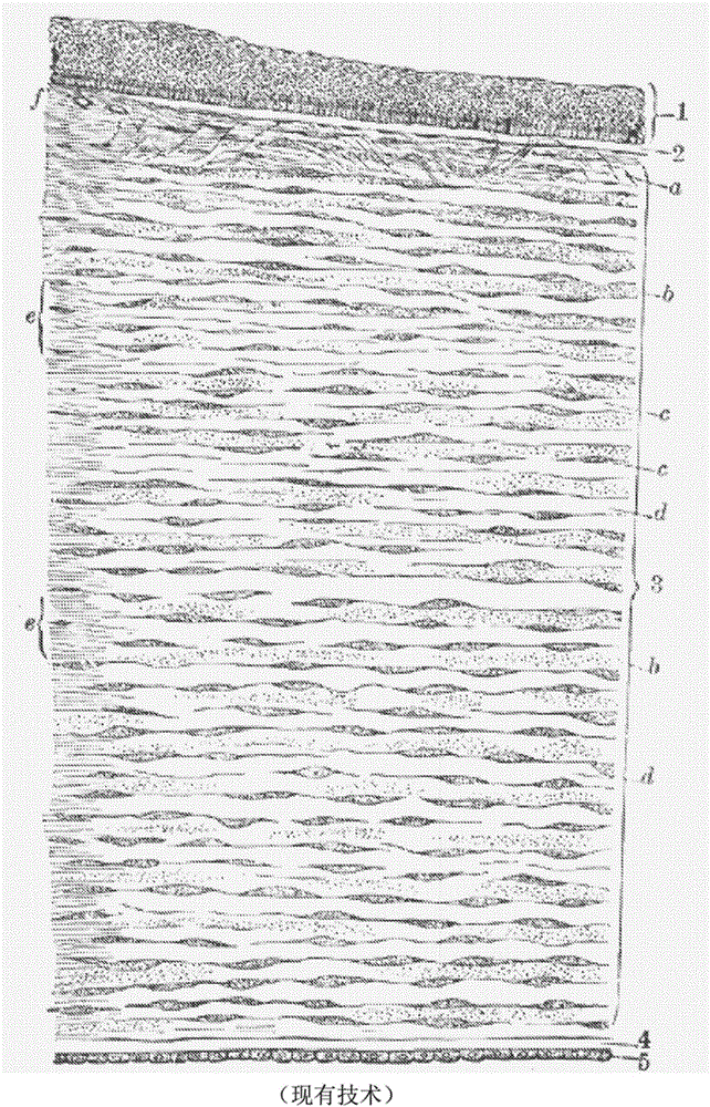
[0078] figure 2 is a longitudinal cross-sectional view (enlarged) of the human cornea near the limbus and shows 1. Epithelium, 2. Bowman's membrane (Anterior elastic lamina), 3. Lamina propria, 4. Bowman's membrane (Desmeek's membrane), 5. Endothelium of the anterior chamber, a. Oblique fibers in the anterior layer of the lamina propria, b. Lamina The fibers of which are cut transversely, creating a punctate appearance, c. the corneal corpuscle, which is fusiform in cross-section, d. the lamina, whose fibers are cut longitudinally, e. the transition to the sclera, It has more pronounced fibrillation, covered by thicker epithelium, and f. small vessels, which incise laterally near the edge of the cornea.

[0079] Embodiments of the present invention are directed to improved devices and techniques for endothelial grafting by techniques such...


PUM


Abstract

A tissue holding assembly for endothelial implantation comprising an open frame having legs for engaging a stem, such that said frame is insertable between a stroma and a Descemet's membrane, said frame being coated with a biological adhesive for adhering to a perimeter of a section of the Descemet's membrane to adhere the section of the Descemet's membrane to the frame for surgical separation of said section by cutting therearound, wherein said frame further comprises a heating element around its inner perimeter.

Description

Background technique [0001] Corneal transplantation is necessary for the surgical treatment of endothelial diseases of the cornea, including glaucoma, edema, and Fuchs endothelial dystrophy. [0002] The cornea is the transparent front part of the eye that contains the iris, pupil, and anterior chamber. The cornea, with the anterior chamber and lens, refracts light and accounts for about two-thirds of the eye's total optical power. [0003] In humans, the refractive power of the cornea is about 43 diopters. Although the cornea contributes most of the eye's focusing power, its focus is fixed. On the other hand, the curvature of the lens is adjusted by the eye muscles to "adjust" the focus of the eye, which is used to bring objects at different distances into focus. [0004] Because transparency is critical, the cornea is avascular and has no direct blood supply, so it receives oxygen that first dissolves in tears and then diffuses to the cornea to keep it healthy. It receiv...

Claims


Application Information

Patent Timeline
Patent Type & Authority Applications(China)
IPC IPC(8): A61F2/14A61F9/01A61F9/007
CPCA61F2/142A61F9/0081A61F2009/00872A61F2/148A61F9/007A61B18/12A61B2018/1407
Inventor 约书亚·本努恩纳坦·戈尔佩林
Owner E K - D D S
Learn More