Ultrasonic-cavitation-injury-based method for establishing atherosclerotic-plaque animal model and blood vessel endothelial injury device
A technology of atherosclerosis and animal models, which is applied in the field of establishing experimental animal disease models, which can solve problems such as increasing the injection dose, reducing the success rate of modeling, and not being easy to model AS plaques, achieving the effect of reducing difficulty and complexity
- Summary
- Abstract
- Description
- Claims
- Application Information
AI Technical Summary
Problems solved by technology
Method used
Image

Examples
example 1
[0048] Instruments and contrast agents:
[0049] FU transducer parameters: single array element FU transducer, center frequency 1.2MHz, focal length 120mm, outer diameter 156mm, middle hole diameter 50mm, acoustic-electric conversion efficiency 72%, half-height width 8mm*1.2mm;
[0050] B-ultrasound imaging probe: Anke AUS-3500 phased array;
[0051] Full digital color ultrasonic diagnostic instrument: Anke AUS-3500;
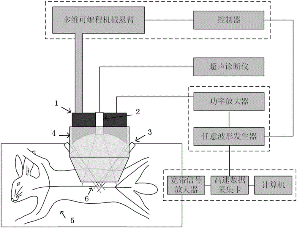
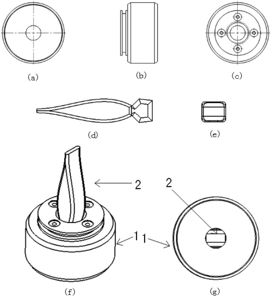
[0052] Multi-dimensional programmable mechanical cantilever: electric three-dimensional mobile platform, including three-dimensional mobile mechanical axis, cantilever and spherical joint. The cantilever is fixed on the mechanical shaft, the spherical joint is fixed on the cantilever, and the probe fixing device 4 is fixed on the spherical joint; the probe fixing device 4 is made of plexiglass and is used to fix the composite probe;
[0053] Power amplifier: AG1016, T&C Power Conversion, USA;
[0054] Arbitrary waveform generator: AWG2021, Sony / Tektronix, JP;...
example 2
[0067] Instruments and contrast agents:
[0068] FU transducer parameters are: 1.1 / 5MHz dual-frequency ring array transducer, outer ring 1.1MHz, inner ring 5MHz, focal length 60mm, outer diameter 103mm, middle hole diameter 25mm;
[0069] B-ultrasound imaging probe: Ultrosonix PA4-2 / 20;
[0070] Full digital color ultrasonic diagnostic instrument: Ultrosonix RP;
[0071] Other equipment is as described in Example 1;
[0072] Contrast agent: microbubble contrast agent.
[0073] Damage implementation steps:
[0074] (1) Anesthetize the rabbit with a respiratory anesthesia machine, prepare the skin on the abdomen, and fix the rabbit on the operating table with the abdomen facing upward.
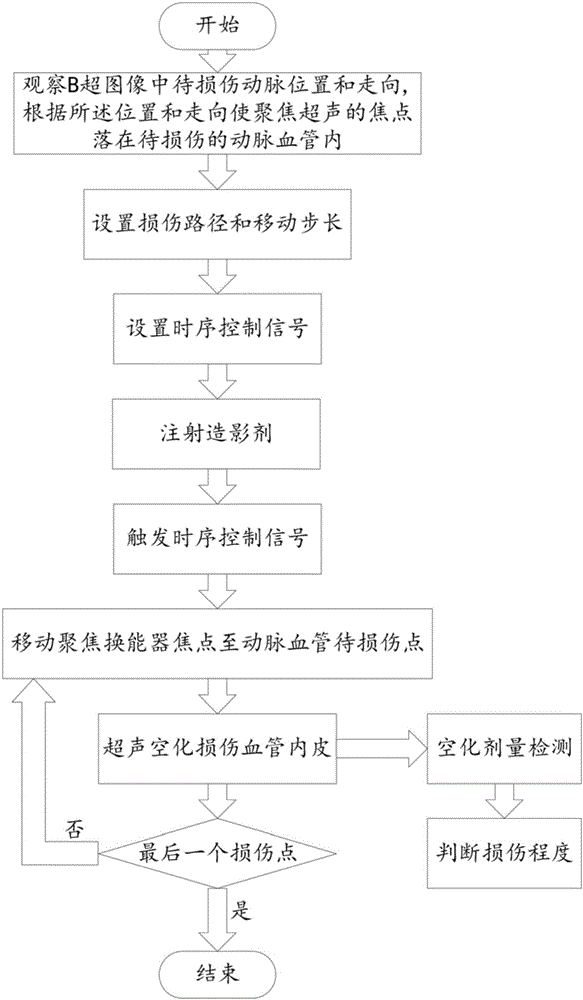
[0075](2) Observe the position and direction of the abdominal aorta to be damaged in the B-ultrasound image, and make the focus of the focused ultrasound fall on the artery to be damaged according to the position and direction.
[0076] (3) Set the path and moving step of the electric three...
example 3
[0080] Instruments and contrast agents:
[0081] Multi-dimensional programmable mechanical cantilever: six-axis robotic arm
[0082] Other equipment is as described in Example 1.
[0083] Contrast agent: microbubble contrast agent.
[0084] Damage implementation steps:
[0085] (1) Anesthetize the rabbit with a respiratory anesthesia machine, prepare the skin on the abdomen, and fix the rabbit on the operating table with the abdomen facing upward.
[0086] (2) Observe the position and direction of the abdominal aorta to be damaged in the B-ultrasound image, and make the focus of the focused ultrasound fall on the artery to be damaged according to the position and direction.
[0087] (3) Set the moving path and moving step of the six-axis manipulator according to the requirements of the direction of the abdominal aorta and the injury area (for example: move 1cm along the direction of the blood vessel, and the step length is 2mm), and load the timing control signal of the arb...
PUM
| Property | Measurement | Unit |
|---|---|---|
| Center frequency | aaaaa | aaaaa |
| Focal length | aaaaa | aaaaa |
| Outer diameter | aaaaa | aaaaa |
Abstract
Description
Claims
Application Information
 - R&D
- Intellectual Property
- Life Sciences
- Materials
- Tech Scout
- Unparalleled Data Quality
- Higher Quality Content
- 60% Fewer Hallucinations
Browse by: Latest US Patents, China's latest patents, Technical Efficacy Thesaurus, Application Domain, Technology Topic, Popular Technical Reports.
© 2025 PatSnap. All rights reserved.Legal|Privacy policy|Modern Slavery Act Transparency Statement|Sitemap|About US| Contact US: help@patsnap.com