
Fluorescence imager and method for imaging lesion area

An imager and lesion area technology, applied in the field of molecular imaging, can solve the problems of reducing the survival rate of patients with chemotherapy effects, the degree of complete removal affecting development, patient health and functional damage, etc., achieving wide selection, small size, and good mobility. Effect

Active Publication Date: 2019-07-30
WUHAN HUAMOU OPTOELECTRONIC TECH CO LTD
View PDF3 Cites 3 Cited by
  • Summary
  • Abstract
  • Description
  • Claims
  • Application Information

AI Technical Summary

Problems solved by technology

The number of cancer cells left after surgical resection is an indicator to measure the success of surgical resection: the number of cancer cells left and the degree of complete removal greatly affect the later development of the disease. If there are too many cancer cells after surgery If the cells remain on the organ, it may directly reduce the effect of subsequent chemotherapy and the patient's chances of survival
[0003] It is difficult for doctors to accurately judge small-sized cancer tissues or tumor borders from naked eyes and touch experience
Moreover, in the patient's lesion area, cancer tissue intersects with normal tissues such as blood vessels and nerves, which cannot be distinguished by naked eyes. Great damage to health and function

Method used


Image

  • Fluorescence imager and method for imaging lesion area
  • Fluorescence imager and method for imaging lesion area
  • Fluorescence imager and method for imaging lesion area

Examples


Embodiment Construction

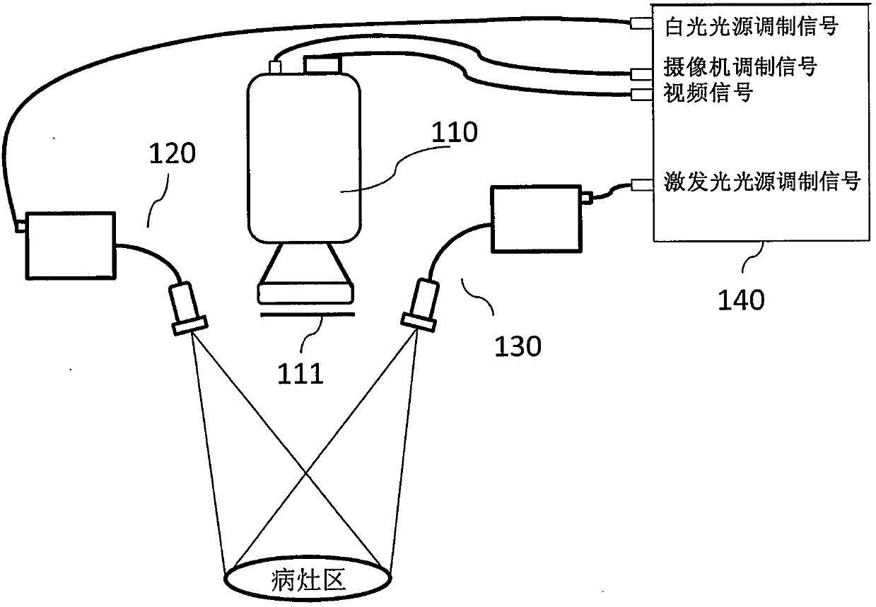
[0029] Most of the current fluorescence imaging systems are usually designed to have two or three cameras work at the same time in order to achieve the ability to simultaneously collect fluorescently labeled images of the lesion area, and divide the work to complete the task of real-time collection of different spectral images, and then the different spectral images Synthetic drug-targeted surgical images.

[0030] Existing fluorescent imaging systems with two or three cameras are bulky, cannot be miniaturized, and have poor mobility. Moreover, two or three cameras work at the same time, and the positions cannot be accurately overlapped, making image synthesis difficult and error-prone.

[0031] What needs to be pointed out is that the fluorescence imaging system with two or three cameras working at the same time must use visible illumination light photography for the whole lesion area, if the fluorescence imaging system in the visible light band is used for some tissues (such ...


PUM


Abstract

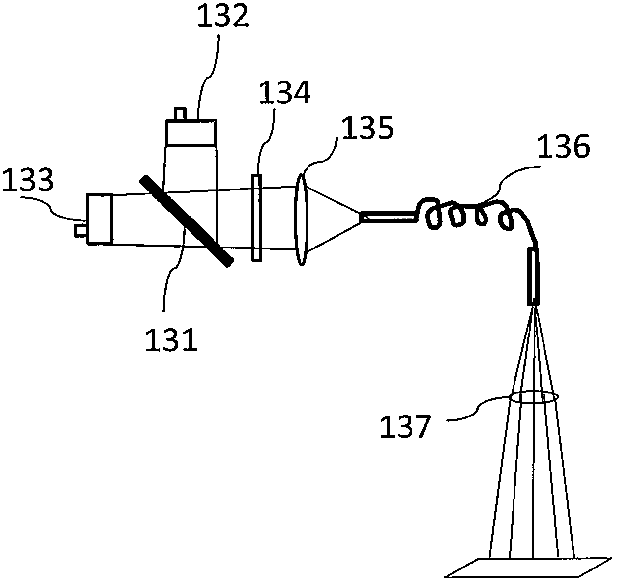
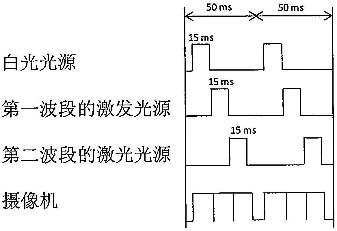
The invention discloses a fluorescent imaging instrument and a focal zone image shooting method, wherein the fluorescent imaging instrument comprises a camera, illuminating light sources and time-sharing multiplexing excitation light sources; the camera is suitable for acquiring images of a focal zone; the illuminating light sources are suitable for providing illuminating light so as to illuminate the focal zone; and the time-sharing multiplexing excitation light sources are suitable for providing at least two exciting light sources differing in waveband; exciting light is configured to excite targeted drugs corresponding to the focal zone to transmit fluorescent light; the exciting light sources differing in waveband share light paths of the time-sharing multiplexing excitation light sources; and the exciting light sources differing in waveband are different in time sequence of exciting the corresponding targeted drugs. According to the embodiment, the fluorescent imaging instrument is small in volume, high in precision degree and is capable of conducting image synthesis on the targeted drugs of the fluorescent light within a visible light waveband; and according to the embodiment, the focal zone image shooting method is high in precision degree, low in interference and is capable of conducting image synthesis on the targeted drugs of the fluorescent light within the visible light waveband.

Description

technical field [0001] The invention relates to the technical field of molecular imaging, in particular to a fluorescence imager and a method for imaging a lesion area. Background technique [0002] The most effective methods of cancer treatment are surgical resection and chemotherapy. The number of cancer cells left after surgical resection is an indicator to measure the success of surgical resection: the number of cancer cells left and the degree of complete removal greatly affect the later development of the disease. If there are too many cancer cells after surgery Cells left on the organ may directly reduce the effect of subsequent chemotherapy and the patient's chances of survival. [0003] For small-sized cancer tissue or tumor border, it is difficult for doctors to make accurate judgments from the naked eye and touch experience. Moreover, in the patient's lesion area, cancer tissue intersects with normal tissues such as blood vessels and nerves, which cannot be dist...

Claims


Application Information

Patent Timeline
Patent Type & Authority Patents(China)
IPC IPC(8): A61B5/00
Inventor 王平
Owner WUHAN HUAMOU OPTOELECTRONIC TECH CO LTD
Learn More